Author: Savannah Spires

  • Microscopic Masterpiece

    This may be bad to admit, but I love a good TikTok scroll. It’s probably not the best use of my time, but I’ve come across some interesting things! New music artists to listen to, events around my city, and even some science accounts. A bit ago, I found a medical student who makes histological artwork and I was absolutely fascinated. Her name is Kait and her paintings are absolutely beautiful (@createbykait on TikTok if you are curious)!

    Of course then I had to do a deep dive about histology. What actually is it? What is she really painting? I assumed it was cells from the look of it, but that was the extent of my knowledge. Turns out there is a lot more to histology than just a pretty picture.

    Histology is the study of microscopic structures of tissues. It usually includes sectioning a thin slice of tissue, staining it with dyes that adhere to different types of structures, then observing the slice under a microscope (NCBI Bookshelf, 2023). Histology is also the foundation for pathology, which is the study of the effects of disease on the body. I had heard pathology as a medical specialty, but again, did not know what they did.

    Turns out, pathologists look at tissue samples from biopsies or autopsies to make diagnoses. I got to watch a brain biopsy earlier this year while shadowing a neurosurgical oncologist. These patients had gotten MRIs that showed a spot of something irregular, called a lesion. During the biopsy, the surgeons took a small sample of the lesion tissue to send to a pathologist to determine what it was, whether it was cancer, an aneurysm, diseased tissue, or even a parasite. The first sample they took is called the frozen section, which they would send to pathology immediately. The pathologists would quickly freeze the sample and examine it to make sure it was usable. The surgeons would have to stay scrubbed in and wait in the operating room until they got confirmation from the pathologist that the sample was good. If it wasn’t, the surgeons would go in and get another sample before closing the wound.

    Pathologists also examine autopsies, or tissue from dead organisms. This is often used when there is an investigation with an unclear cause of death. This led me to Marianne Hamel, a forensic pathologist, who recognized how paradoxically beautiful these histological images can be. So much so that she created a collection of histological images that is now on display at Rudger’s University (Hamel & Johnson, n.d.). She said in an interview that she hopes to educate people about her career field, but also allow people to appreciate the beauty of the human body (Chandler, 2023).

    Histology has evolved a lot since its beginnings. Hamel said photos used to be shot in black and white, making interpreting the images much more difficult. Now there is a plethora of different dyes used to make multicolored images. This offers so much more information for pathologists to gather, but also a higher level of difficulty. Learning to read [histological] slides, Hammel says, is like learning another language (Chandler, 2023).

    It’s funny how what seems like just a pretty picture is actually such an intricate science and can show how beautiful life can be.
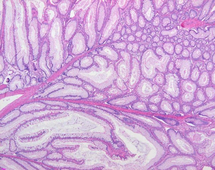
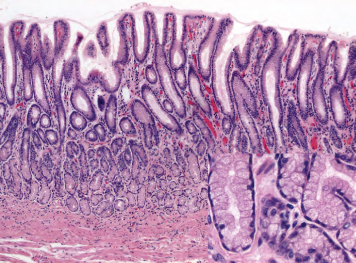
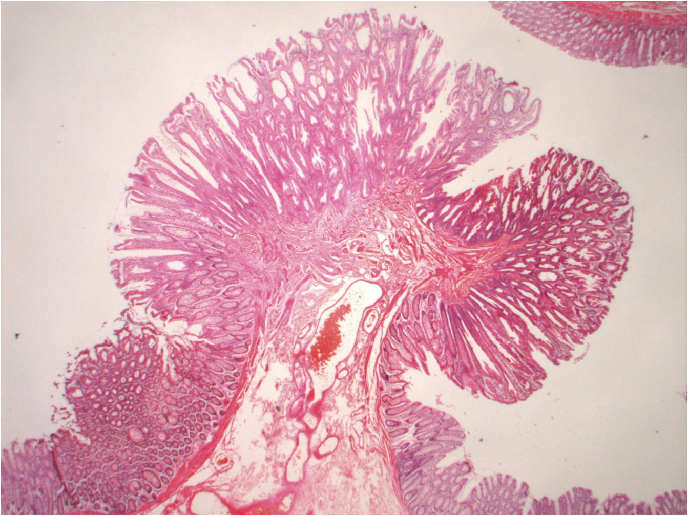
    “Mixed histology polyp.” Image by Caoimhin74, via Wikimedia Commons, licensed under CC BY-SA 3.0.
    “Histology of normal antral mucosa” Image by Carrasco G, Corvalan AH, via Wikimedia Commons, licensed under CC BY-SA 3.0.
    “Mixed histology polyp” Image by Caoimhin74, via Wikimedia Commons, licensed under CC BY-SA 3.0.

    References

    National Center for Biotechnology Information. (2023). NCBI Bookshelf. U.S. National Library of Medicine. https://www.ncbi.nlm.nih.gov/books/NBK557663/

    Hamel, M., & Johnson, N. (n.d.). Death under glass. https://www.deathunderglass.com/

    Chandler, R. (2023, October 31). Post-mortem histology artwork brings death to life. Evident Scientific. https://evidentscientific.com/en/insights/post-mortem-histology-artwork

  • Filling in the Gaps: Biases and Pareidolia

    Our brains are amazing, allowing us to do so much. Even so, they aren’t perfect. Do you remember during COVID, you would meet people for the first time wearing masks? Then, when you finally saw their whole face, you were surprised. And it’s not like you had purposefully thought about what the bottom half of their face looked like, but it was still somehow different than expected.

    Facial Recognition & Pareidolia

    This may be due in part to the fact that humans are wired to recognize faces. This is such a strong skill that we even recognize “faces” in inanimate objects: the front of cars, electrical outlets, even clouds or rocks. This phenomenon is called pareidolia, a human tendency to recognize meaningful patterns, especially faces. This is thought to evolve from the need to quickly recognize facial expressions for threats or social cues. We even have an entire region of the brain responsible: the face fusiform area (FFA) (Zhou & Meng, 2020). Studies have shown that even infants can recognize face-like patterns as faces (Kato & Megitani, 2015). Infants as young as seven months were able to recognize faces in Arcimboldo’s paintings (Kobayashi et al., 2012). These paintings were of inanimate objects, such as vegetables, arranged to resemble a portrait of a human. They are honestly a bit creepy, but feel free to give it a google!

    Obviously we can all recognize a face, even under a mask, but a study using an artificial face recognition system gave us some evidence for why. A system called deep convolutional neural networks, or DCNN, determined the most important facial feature for facial recognition to be the eyebrows. This was followed by the eyes, mouth, then nose (Zhang et al., 2024). This makes sense with facial recognition under a mask, as the two most important features are still visible. Why the eyebrows are the most important isn’t clear. It could be that eyebrows are important for facial expressions, or that they are important for individual recognition due to their large diversity in color, shape, and placement.

    Cognitive Dissonance & Bias

    I found the science behind facial recognition fascinating, but it still doesn’t quite explain why we subconsciously fill in the bottom half of someone’s face. As it turns out, recognizing parts of a meaningful pattern, such as the face, but missing key parts of it, creates cognitive dissonance. Cognitive dissonance is when there is discrepancy between what your brain expects or believes and the information it is gathering. In the case of a mask, your brain is expecting to see all the important components of a face, so when the mouth and nose are missing, it creates a sort of tension. To relieve this tension, your brain uses what it knows about the face to fill in the gaps. This way, the pattern fits in with the categories we already have.

    Significance

    Pattern recognition, especially facial recognition, is super important, allowing us to recognize slight changes in facial expression or obstructed faces, like under a mask. But this can also lead to something not so great. Amy E. Herman said in her book “Visual Intelligence” that humans see through the brain, not the eyes. All your sensory organs collect signals and send them to the brain for processing. The signals don’t mean anything on their own, whether that is light through the eyes or smell through the nose. It is the processing that allows us to process these signals into something meaningful. This information is then tried to fit in with our prior knowledge, reducing cognitive dissonance. But in the process of fitting in new information, it can get twisted. Filling in what a face looks like under a mask, for example, is the brain twisting information to fit it in with what it already knows. This phenomenon happens all the time and is the science behind confirmation bias.

    Human brains are extremely susceptible to confirmation bias, even going so far as to discard or ignore information that contradicts our beliefs (The science of stereotyping, 2022). The Smithsonian has a super cool interactive article about the science of bias if you are interested in learning more!

    All of this to be said, our brains are amazing things, but they are not perfect. It is important to know that sometimes they take the easy path, twisting information to fit what we already know, from what someone looks like under a mask, to knowing a face-shaped rock is not a person.


    References

    Zhou, L.-F., & Meng, M. (2020). Do you see the “face”? Individual differences in face pareidolia. Journal of Pacific Rim Psychology, 14, e2. https://doi.org/10.1017/prp.2019.27

    Kato, M., & Mugitani, R. (2015). Pareidolia in infants. PLOS ONE, 10(2), e0118539. https://doi.org/10.1371/journal.pone.0118539 

    Kobayashi, M. et al. (2012). Do infants recognize the Arcimboldo images as faces? Behavioral and near-infrared spectroscopic study. Journal of Experimental Child Psychology, 111(1), 22-36. https://doi.org/10.1016/j.jecp.2011.07.008. 

    Zhang, Q. et al. (2024). Understanding of facial features in face perception: insights from deep convolutional neural networks. Frontiers in Computational Neuroscienc, 18 https://doi.org/10.3389/fncom.2024.1209082 

    University of Wisconsin–Madison Department of Psychology. (2022). The science of stereotyping. https://psych.wisc.edu/news/the-science-of-stereotyping/

    Smithsonian Institution. (2021). The science of bias. https://biasinsideus.si.edu/online-exhibition/the-science-of-bias

  • Secrets Of The Skin: A Hidden Ecosystem

    Every system in our body is connected, and every process in it is affected by all others. When two processes affect each other in a bidirectional manner, they are called axes. Scientists have recently looked into the gut-skin-brain axis, or the connection between your gut and skin health and your psychological well-being.

    It is commonly known that your gut has its own microbiome, consisting of bacteria that help us digest our food. Less people know that there is a similar microbiome on your skin. Right now, there are thousands of different species of bacteria, fungi, viruses, and other microorganisms living in symbiosis on the surface of your skin. The word microbe usually comes with a negative connotation, making you think of disease or parasites, but the microbes both on our skin and in our gut are vital to our health. Having an unbalanced gut microbiome, or being in dysbiosis, is often associated with various skin conditions, such as eczema, psoriasis, rosacea, or acne (Wiginton 2025).

    The skin makes for a very harsh environment. It is dry and we are constantly shedding skin cells, which makes it a less than ideal place for a microbe to live. But many species have adapted to be able to handle life on the skin, and most have preferred locations on the body as well. Some bacteria prefer somewhere warm, such as the armpit or foot, whereas others thrive somewhere oily, like our face. These microbes actually work as part of our immune system. Our skin is our largest organ and prevents pathogens from entering our body. The microbes that thrive on our skin like the harsh environment, and help keep it so. Other harmful microbes often cannot survive in these conditions, and sometimes are even attacked by our skin’s microbes (Byrd et al. 2018).

    The link between the gut microbiome and our psychological well-being has been well established as bidirectional, meaning they affect each other. Dysbiosis, or an imbalance, in the gut has been linked to depression, anxiety, and autism. On the other hand, stress can alter our gut microbiome and make us more susceptible for gastrointestinal disorders. Studies have even shown which bacteria, Cutibacterium, show the strongest link to our psychological well-being (Tyson-Carr et al. 2025).

    Recently, proof has been found of a triple axis, the gut-skin-brain axis. Probiotics are living microorganisms in our gut and have been found to play a part in our immune system. These microbes in the gut help our immune cells distinguish between harmful and beneficial microorganisms. Bacteria in the gut also release molecules that have anti-inflammatory effects on the skin. Studies have shown that taking probiotics benefits your gut microbiome, which in turn benefits our immune system, creating a snowball effect that can be beneficial towards our skin health and psychological well-being. Probiotics have even been proven to improve some chronic inflammatory skin diseases, such as psoriasis by increasing certain microbe species in the gut (Abdi et al. 2024).


    Wiginton, K. (2025). What Is the Skin Microbiome? WebMD. https://www.webmd.com/skin-problems-and-treatments/skin-microbiome

    Byrd, A. L., Belkaid, Y., & Segre, J. A. (2018). The human skin microbiome. Nature Reviews Microbiology, 16(3), 143–155. https://www.nature.com/articles/nrmicro.2017.157

    Tyson-Carr, J., Leng, J., Scott, M., Adams, S., Hoptroff, M., Murphy, B., Fallon, N., Paterson, S., Thomas, A., Giesbrecht, T., & Roberts, C. (2025). Body site-specific associations between human skin microbiome composition and psychological wellbeing. British Journal of Dermatology, 193(Supplement 2), ii6–ii14. https://academic.oup.com/bjd/article/193/Supplement_2/ii6/8140349

    Abdi, A., Oroojzadeh, P., Valivand, N., Sambrani, R., Lotfi, H. (2024). Immunological aspects of probiotics for improving skin diseases: Influence on the Gut-Brain-Skin Axis. Biochemical and Biophysical Research Communications, 702. https://www.sciencedirect.com/science/article/pii/S0006291X24001682


  • What’s Making That Smell? The Chemistry Behind Common Scents

    The Science of Smell

    Of the five senses, scientists understand the least about smell. We recognize scents when small airborne molecules stimulate receptors in the back of our nose, which then send signals to our brain. Humans have about 500 different types of receptors, each one specific to a different scent molecule (“How the nose decodes complex odors”, 2020).

    But what makes some molecules have scents? Well, the molecules must be able to travel through the air to the back of the nose, so they have to be volatile. Volatility is how easily the molecule evaporates at room temperature. The more a molecule evaporates into the air, the more likely it is that it reaches the receptors in the back of our nose. Even then, there must be a receptor for that specific molecule for it to be recognized (Gang, 2005).

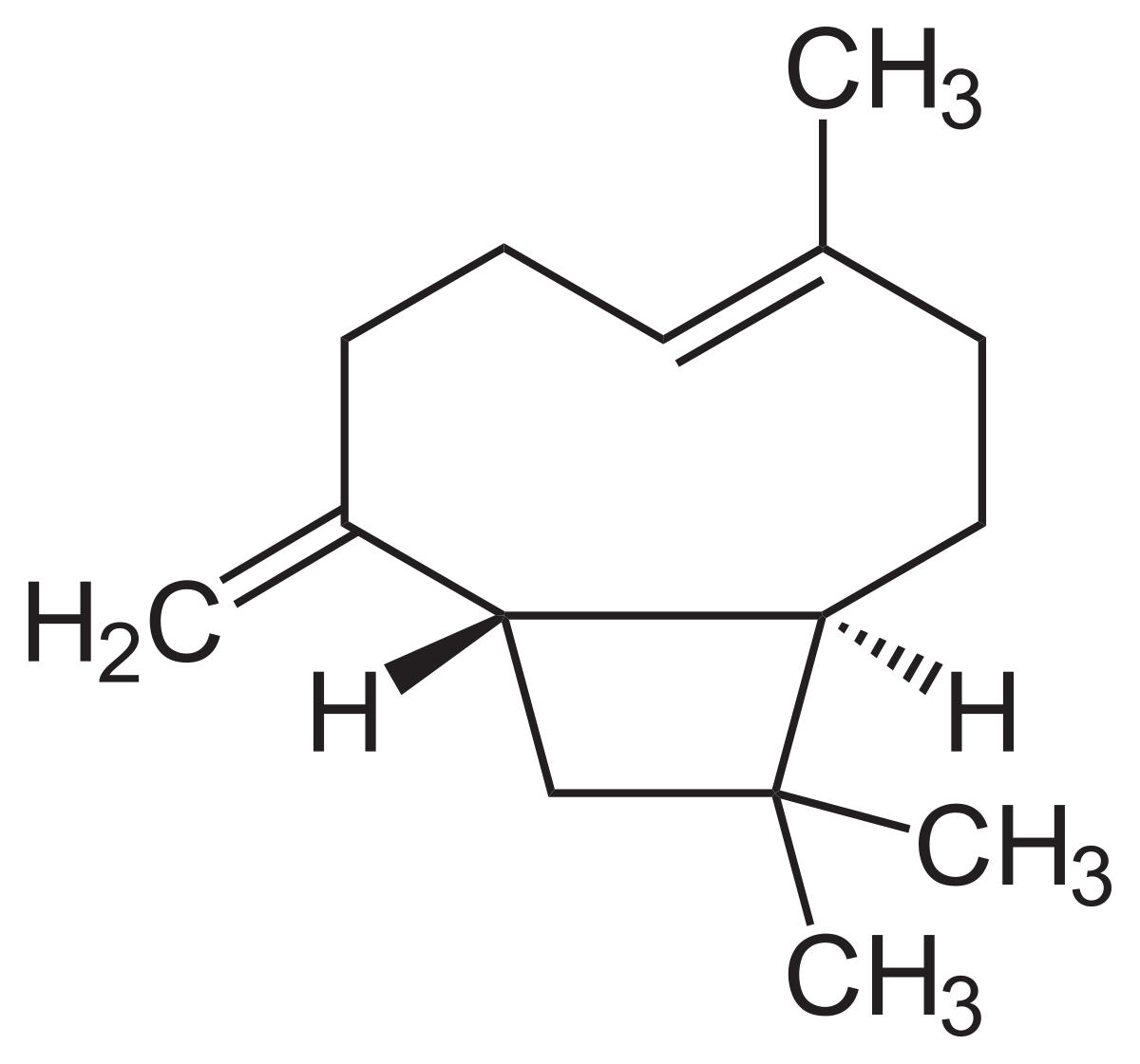
    The whole process seems simple enough, but most scents are a combination of tens to hundreds of different scent molecules. The smell of chocolate and cocoa products, for example, is made of over 600 scent molecules (Counet et al., 2002). Scientists still don’t fully understand how more complicated scents work, (Gottfried & Wilson, 2011) but most have been found to be made of just a few major compounds, such as the southern magnolia flower. Its scent comes from a mixture of 14 different molecules, but two, beta-caryophyllene and beta-cedrene, are the main compounds (Garg & Kumar, 1999).

    Beta-caryophyllene
    Beta-cedrene

    Many common scents can be traced back to specific molecules. Even small changes in a molecule can cause large changes in its scent. Check out a few familiar scents below!

    Common Scent Molecules

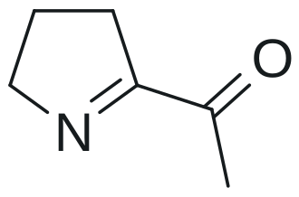
    6-Acetyl-2,3,4,5-tetrahydropyridine gives popcorn and baked goods their typical smell. A slight change creates 2-Acetyl-1-pyrroline, which is the main component of rice’s smell (Dhrubo, 2015).

    6-Acetyl-2,3,4,5-tetrahydropyridine
    2-Acetyl-1-pyrroline

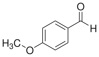
    4-Anisaldehyde is used a lot in the fragrance industry. It has a very strong sweet floral scent and is very similar to vanillin. Vanillin, as you may have guessed, is the main component of vanilla’s scent. Natural vanilla extract is made of hundreds of different molecules, but synthetic vanilla scent is usually just pure vanillin (Dhrubo, 2015).

    4-Anisaldehyde
    Vanillin

    Benzaldehyde has an almond-like odor and is very useful in the fragrance industry because of its simple structure (Dhrubo, 2015).

    Benzaldehyde

    Cinnamaldehyde makes up for around 90% of the scent of cinnamon (Dhrubo, 2015).

    Cinnamaldehyde

    Natural honey is an extremely complicated scent, made up of 187 volatile compounds, and varies based on its nectar source (Mahmoud et al., 2024). Phenylacetaldehyde is the main component of honey’s scent, but methyl phenylacetate is used for a synthetic honey scent because of its strength. It only takes a little bit of methyl phenylacetate to make a perfume or candle honey-scented (Dhrubo, 2015).

    Phenylacetaldehyde
    Methyl Phenylacetate

    Even fresh cut grass has its own molecule. (Z)-3-hexenal is one of the molecules released when grass is damaged, and is what gives it that freshly mowed lawn smell (Dhrubo, 2015).

    (Z)-3-Hexenal

    If you want to learn more about fun molecules, the American Chemical Society (ACS) has been publishing a “Molecule of the Week” since 2001. This past week’s molecule was myristicin, which is a primary component of many spices such as nutmeg, dill, parsley, and anise. Interestingly, its structure is very similar to that of amphetamines and can cause hallucinations if ingested in high doses (much higher than used for cooking). On their website, they have an archive of over one thousand molecules, complete with properties, history, and fun facts! Click here to check it out.


    How the nose decodes complex odors. (May 12, 2020). National Institute of Health. https://www.nih.gov/news-events/nih-research-matters/how-nose-decodes-complex-odors

    Gang, D. (June 2005). Evolution of Flavors and Scents. Annual Review of Plant Biology. 56, 301-325. https://www.annualreviews.org/content/journals/10.1146/annurev.arplant.56.032604.144128

    Gottfried, J., Wilson, D. (2011). Neurobiology of Sensation and Reward. National Library of Medicine. https://www.ncbi.nlm.nih.gov/books/NBK92786/

    Counet, C., Callemien, D., Ouwerx, C., Collin, S., (March 15, 2002). Use of Gas Chromatography−Olfactometry To Identify Key Odorant Compounds in Dark Chocolate. Comparison of Samples before and after Conching. Journal of Agricultural and Food Chemistry, 50(8), 2385–2391. https://pubs.acs.org/doi/10.1021/jf0114177

    Garg, S., Kumar, S., (1999). Volatile constituents from the flowers of Magnolia grandiflora L. from Lucknow, India. Journal of Essential Oil Research 11(5),, 633-634. https://aromadb.cimapbioinfo.in/web_essentialoil_details.php?id=CRESOL159

    Mahmoud, M., Kilic-Buyukkurt, O., Aboul Fotouh, M., Selli, S. (October 2024). Aroma active compounds of honey: Analysis with GC-MS, GC-O, and molecular sensory techniques. Journal of Food Composition and Analysis, 134(106545). https://www.sciencedirect.com/science/article/pii/S0889157524005799#:~:text=Compounds%20such%20as%20phenylacetic%20acid,the%20distinctive%20smell%20of%20honey.

    Dhrubo, J. S. (2015) Esters, Terpenes And Flavours: Make The Mood Cheers by Three Musketeers!. World Journal of Pharmaceutical Medicine, 4(8), 1-40. https://wjpr.s3.ap-south-1.amazonaws.com/article_issue/1438667348.pdf

  • Multicellular Archaea?

    All life is classified into three overarching domains: archaea, bacteria, and eukarya. We are all familiar with eukarya, seeing that humans fall under this domain, and bacteria, but archaea are often forgotten. They are defined as single-celled prokaryotes, meaning they exist as a cell without a defined nucleus. Archaea inhabit extreme environments, such as hydrothermal vents or salt lakes, and highly acidic or alkaline conditions. This is the main reason there is not as much information on archaea as there is on bacteria and eukarya. It is difficult to isolate these species from such environments and recreate the conditions in a lab setting.

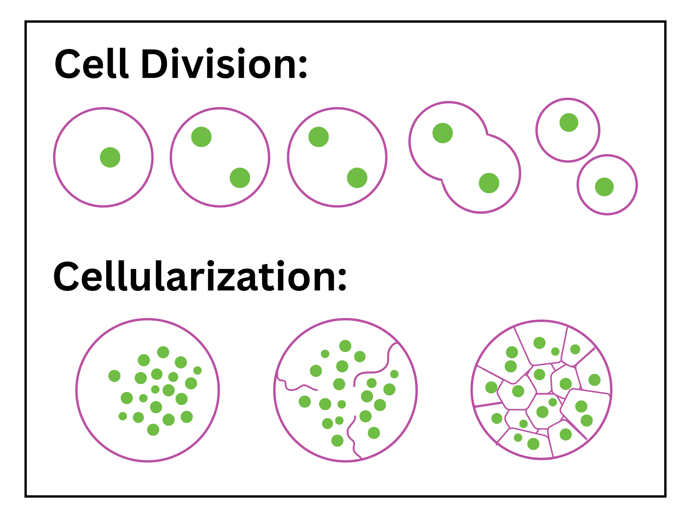
    But new findings in cell biology have put the single-cellularity of archaea into question. Normally, when under excessive pressure, cells will halt all cell division as a protective mechanism to ensure all cells have adequate volume and are without genetic defects. Recently, a species of archaea found in hypersaline environments, H. volcanni, was found to undergo a process closely resembling cellularization instead of regular cell division when under pressure. Cell division is the more common and orderly process, usually mitosis and cytokinesis, where a single cell replicates its DNA once and increases in size until it splits into two daughter cells. Cellularization, on the other hand, is when a single cell replicates its DNA until it has multiple nuclei, then splits into many smaller cells. Archaea do not have nuclei, but the process they underwent in the experiment strongly resembled that of eukaryotic cellularization.

    In animals, cellularization is rare, primarily found in the early development of fruit fly embryos. In this case, the process forms tissues, which are the basis of multicellular organisms, defined as a group of cells that work together for a common function. The multicellular archaea can’t quite be defined as tissue, since they don’t have the connection and interaction that tissue does, but it is definitely an interesting new find. This also serves as evidence for the theory that multicellularity evolved before the teamwork seen in multicellular organisms.

    H. volcanii also has certain traits that make this process favorable. They naturally contain multiple copies of their DNA spread throughout the cell, and their cell division is influenced by the pressure of their surroundings, which they recognize through their membrane tension. The cellularization also produces two types of cells, peripheral and central scutoid cells, each with distinct structural protein patterns. Further implications of this are unknown, but it will be exciting to watch this newfound trait of archaea unfold.

    Pillai, E., & Brunet, T. (2025). Archaea go multicellular under pressure. Science, 388(6742), 28-29. https://www.science.org/doi/10.1126/science.adw6689?utm_source=sfmc&utm_medium=email&utm_campaign=ScienceAdviser&utm_content=distillation&et_rid=1095835014&et_cid=5579756
    Niederberger, T., & et al. (1998). Archaea. Encyclopedia Britannica, https://www.britannica.com/science/archaea
    Rados, T., Leland, O., Escudero, P., Mallon, J., Andre, K., et al. (2025). Tissue-like multicellular development triggered by mechanical compression in archaea. Science, 388(6742), 109-115. https://www.science.org/doi/10.1126/science.adu0047#:~:text=The%20advent%20of%20clonal%20multicellularity,at%20a%20critical%20cell%20size
    Liu, C. (2017). Archaea. Innovative Genomics Institute (IGI). https://innovativegenomics.org/glossary/archaea/

  • Hidden Vegetables & Essential Vitamins

    We all know vitamins are important, but what do they actually do? Well, each vitamin has different benefits, with some playing multiple roles in important bodily functions. The oxygen transfer from our lungs to our cells, for example, depends on many vitamins and minerals, such as vitamin B6, C, and iron.

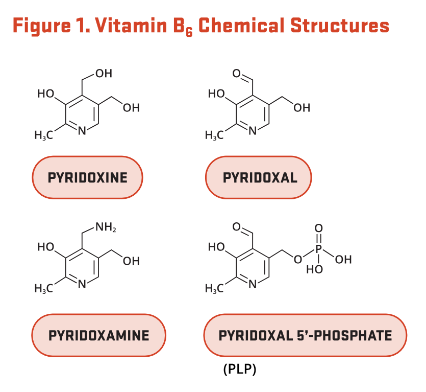
    Vitamin B6 comes in many different forms, but our bodies convert it to its most useful one: pyridoxal-5′-phosphate, conveniently referred to as PLP. PLP is a coenzyme that helps catalyze the production of hemoglobin.

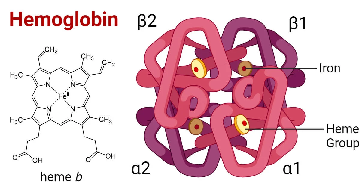
    Found in our red blood cells, hemoglobin is arguably one of the most important molecules in our body. Each hemoglobin molecule contains four heme groups, which are responsible for carrying the oxygen our body needs. Heme groups are also coordination complexes, meaning they have one or more molecules bound to a metal center, which in this case is iron.

    As you might have guessed, this is why iron is so important in our diet. Without it, our body can’t make as much hemoglobin, and our cells wouldn’t get the oxygen they need. This makes sense if you have ever experienced low iron; your cells aren’t getting the oxygen they need to produce energy, which causes fatigue.

    Iron, like vitamin B6, comes in many forms, but there is only one form useful to our body. Iron must have a 2+ charge to form a heme group. The iron we get from meat already has this 2+ charge, but the iron from vegetarian sources often doesn’t. Our body must convert the iron into its usable form, which is where vitamin C comes in.

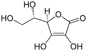
    Vitamin C, also known as ascorbic acid, has numerous jobs to keep our body functional, one of which is helping give iron its 2+ charge so it can form a heme group. It is hypothesized that vitamin C increases the acidity of our digestive tract, making it easier for our body to convert iron into the form we need. This is why patients with iron deficiency are often told to take their iron supplements with a glass of orange juice.

    All together, vitamin B6, C, and iron ensure our cells get the oxygen they need by playing a part in different stages of hemoglobin production. Getting all of the vitamins your body needs can be tricky, but there are many ways to make it happen, especially if you like to bake. Including “hidden” vegetables in meals or desserts is a fun way to do this and can increase your vitamin intake without drastic changes to your diet.

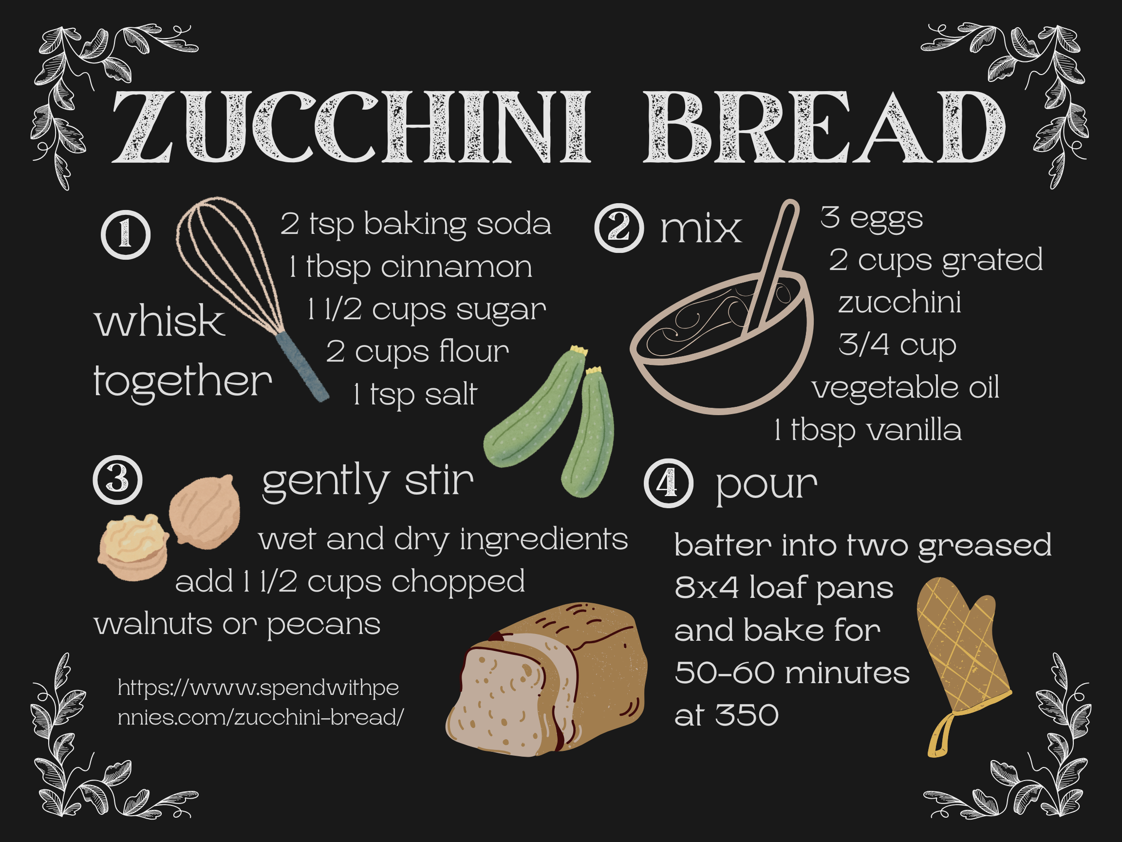
    Since zucchinis contain vitamins B6 and C and nuts are a vegetarian source of iron, this recipe for zucchini bread provides you with three essential vitamins for hemoglobin production along with a delicious breakfast or snack. The zucchini keeps the bread moist without taking away from the warm cinnamon flavor, and the slices taste especially good toasted with butter. Enjoy!

    Dahal, P. (2025, March 6). Hemoglobin decoded: Key types, functions & disorders. Microbe Notes. https://microbenotes.com/hemoglobin/
    Deng, J., & Ramelli, L., & Li, P. Y., & Eshaghpour, A., & Muti Scheunemann, G., & Crowther, M. (2023, November 2). Efficacy of Vitamin C with Iron Supplementation in Iron Deficiency Anemia Patients: A Systematic Review and Meta-Analysis. Ash Publications. https://ashpublications.org/blood/article/142/Supplement%201/1091/504755/Efficacy-of-Vitamin-C-with-Iron-Supplementation-in

    Higdon, J., & Drake, V., & Delage, B. (2024, May). Vitamin B6. Oregon State University. https://lpi.oregonstate.edu/mic/vitamins/vitamin-B6#:~:text=PLP%20functions%20as%20a%20coenzyme,to%20microcytic%20anemia%20(3).

    Wikipedia contributors. (2025, March 22). Heme. Wikipedia, The Free Encyclopedia. https://en.wikipedia.org/wiki/Heme

    Wikipedia contributors. (2025, March 31). Chemistry of ascorbic acid. Wikipedia, The Free Encyclopedia. https://en.wikipedia.org/wiki/Chemistry_of_ascorbic_acid