Tag: neuroscience

  • All Work and No Play

    The cover image was sourced from https://doi.org/10.1016/j.cub.2014.09.020.

    If you’ve ever fallen down the easily addictive, low-stress rabbit hole that is the world of wonderfully awesome animal videos, you might have come across this video of a crow in Russia, sliding down a snow-covered roof while perching on a jar lid.

    While it’s easy to think that this crow went sledding purely for their own enjoyment, scientists aren’t content with that conclusion – thinking in such an anthropomorphic way projects our own human thoughts and feelings onto animals who, while exhibiting familiar behaviors, may not be doing it for the same reasons we do (Emery & Clayton, 2015).

    But, regardless of what we may think, birds do play! What does that look like? Well, most forms of play are divided into one of several categories: the first broad category being locomotor play, which includes all kinds of flight-related play – hanging from surfaces, flying upside down (Emery & Clayton, 2015), and even sledding down a snowy roof in Russia!

    Social play involves interactions between birds – there’s play chasing, where one bird follows another, whether on the ground or through the air. We also have play fighting, where two birds engage in actions adopted from antagonistic behaviors. There’s also play invitation, where two birds engage in actions that occur primarily before play. Finally, we have social object play, where two birds engage with one or more objects in their environment (Bond & Diamond, 2003).

    While we briefly touched on it, object play itself can be considered another major category of play – involving exploration, curiosity, object manipulation, where birds learn about the structure of objects, if they’re edible or not, and how they might work (Emery & Clayton, 2015).

    Figure 2 Source: https://corvidresearch.blog/wp-content/uploads/2015/03/northwestern-crow-youngsters-playing-tug-of-war_595.jpg

    The problem with researching play is that birds don’t necessarily need a reason to play – even when considering the anthropomorphic projection we cast onto them from our human perspectives, it can be as simple as having fun!

    “Fun”, at least, in the context of the brain, involves taking part in something rewarding that provides a sense of pleasure. You may have heard of the neurotransmitter dopamine… it plays an important role in the reward-seeking system, controlling the search for reward-inducing stimuli for almost all creatures (Emery & Clayton, 2015).

    When people come across phenomena like this in the wild, the answer that tends to come to mind often lies on the justifying end of the spectrum; in our attempt to be objective, we may implicitly assign a “value” to that act of play. Perhaps birds play fight to practice hunting, perhaps birds engage in social play to form strong, interconnected networks, and perhaps that crow slid down that roof to learn how to use that lid!

    Studies done on the potential concrete benefits of play – such as relative brain size, for instance – ultimately find that there is little to no predictive value between the complexity of play with those benefits. As such, it is hard to say whether or not the act of play is utilitarian in nature (Bond & Diamond, 2003).

    Does that mean that play is inherently fun? Once again, it’s hard to say! Humans, after all, may be the only species that have the luxury of leisure time. The time we spend solely pursuing pleasure: games, hobbies, activities we may find fun, are all a product of modern developments. We no longer have to hunt or grow our own food, we no longer have to worry about predators, we no longer have to worry about forming civilized societies – but birds, and by extension, all other animals, do (Emery & Clayton, 2015)!

    Perhaps that is the underlying reason why we believe play has to have a purpose. Wild animals have a limited time to find food, find water, avoid predators, find a mate, raise offspring, all in the name of the cycle of life. Why, then, would a bird waste its time playing for the sake of playing? Why, then, do we have examples of crows sledding through the snow?

    All of these questions are interesting to ponder, and I personally don’t have all the answers. The realm of neuroscience is far out of my understanding, but I hope I’ve done a good job setting the stage for this interesting question.

    I’ll leave things here with a plan of action to you, the reader – whenever you see a cute animal video in the future, think to yourself: why could this be happening? Is it play, or is it intentional? Most importantly, though… why is it so gosh darn adorable?!

    Reference List

    Bond, A., & Diamond, J. (2003). A Comparative Analysis of Social Play in Birds. Behaviour, 140(8-9), 1091–1115. https://doi.org/10.1163/156853903322589650

    Emery, N., & Clayton, N. (2015). Do birds have the capacity for fun? Current Biology, 25(1), R16–R20. https://doi.org/10.1016/j.cub.2014.09.020

  • Painting With all the Colors of the Wind Part Two

    PART TWO: WHY DOES SYNESTHESIA HAPPEN?

    So, we know a bit about what synesthesia is. But what actually causes these remarkable perceptions?

    Well the truth is, as usual, that no one really knows for sure. There is strong evidence for a genetic component to synesthesia, as it tends to run in families. Despite this, however, it isn’t inherited directly- that is,  the type of synesthesia, in terms of what stimuli will produce what sensory responses, doesn’t seem to be inherited at all. If your mother had lexical-gustatory synesthesia, for example, you are statistically more likely to have some form of synesthesia, but there is no  preference towards synesthesia involving vision or taste. This, along with the fact that many people have more than one type of synesthesia, suggests that all forms of developmental synesthesia share some common cause and genetic predisposition (Ward). What this genetic component actually does is still contentiously debated though.

    Investigations into synesthesia have been going on for many years. In 1690, John Locke wrote about a blind man who described the color scarlet as “the sound of a trumpet”, and the first well-documented case of synesthesia dates all the way back to 1812, when an Austrian doctor named Georg Sachs included descriptions of his own synesthesia in his medical dissertation (Ward). When the scientist Cornaz reviewed Sach’s paper again in 1848, it generated a lot of interest in the phenomena, and many more cases of synesthesia were discovered and investigated. Cornaz, however, thought that the color-grapheme synesthesia Sachs experienced was related simply to his eyes. He suggested that it was a result of having more color receptors in the eye, and therefore being able to detect additional colors that others couldn’t see (Ward). This explanation has been widely rejected, but it did spark the first significant interest in synesthesia, and people began to suggest other possibilities, such as Lusanna’s 1873 proposal that the “cerebral organs” for music, language and color might be close to each other and might overlap or connect in people with synesthesia (Ward).

    Interest in synesthesia seems to have waned during the early 1900s, but it was definitely revitalized in the 1980s, after the neurologist Richard Cytowic published several case studies and a popular book, “The Man Who Tasted Shapes”, about synesthesia. Although his suggestion that synesthesia might be related to the limbic system has also been discredited, he played a huge role in bringing attention back to synesthesia and starting a new era of neuroscientific and psychological research into it.

    In terms of the current research into the neural mechanism for what is happening in the sensory systems of synesthetes, there seem to be two major competing schools of thought. One views synesthesia as a result of structural differences in the brain and nervous system, and the other claims that it is due to functional differences in use of the neural circuits we all have. To break down each of these hypotheses, we can look a bit more closely into the arguments of each:

    Team Structural Differences
    As the researcher Baron-Cohen sees it, synesthesia is best explained by a difference in the number of connections in the brain- more and unusually strong neural pathways could allow for a physical connection between areas of the brain that typically respond to different senses/stimuli. It has also been suggested, by Dr. Daphne Maurer, that everyone is born with these additional connections, but that they are typically lost are we grow. Under this proposal, synesthesia is therefore a result of changes in development that neglect to “prune” these connections (Carpenter).

    There is data to support the idea that the brains of people with synesthesia are physically different than those without it. Among the many imaging studies that have been done,  a study by Weiss and Fink 2009 found that the intraparietal sulcus had more gray matter in people with color-grapheme synesthesia, and that it is also activated when those participants were viewing graphemes. In addition, imaging data from Rouw and Scholte 2007 found that people with color-grapheme synesthesia had significantly increased white matter connectivity in brain regions they believe to be implicated with synesthesia.


    Team Functional Changes
    Some scientists think that synesthesia is better explained as the result of using the same brain architecture and connections, but in a different way. Proponents of this hypothesis focus on neural pathways common to everyone, but generally inhibited, or blocked, in one direction. Having that pathway be disinhibited would then allow people with synesthesia to distribute information between these neurons differently (Carpenter, Grossenbacher and Lovelace 2001).

    For synesthesia, the major pathways considered are those that function with our sensory systems. A simplified version of these networks goes something like this: Information is carried from receptor cells across the body to different regions of the brain, where it is processed, put into context with the activity of other neurons, and used to form a comprehensive picture of the situation. Then, once the input has been consolidated, and a conscious perception or decision about the course of action has been made, the brain can send feedback and further instructions to the appropriate areas in what are called feed-backward connections.

    The idea, then, is that in synesthesia, the inhibitory nature of some feed-backward connections of the sensory systems is deactivated or “disinhibited”, which means that information may be flowing down paths and/or in directions it normally couldn’t.  This would allow a stimulus to interact with the system differently, and potentially “mix” information from multiple sensory inputs or areas, causing synesthesia (Carpenter).

    Grossenbacher, a lead advocate for this approach to looking a synesthesia, claims that this explanation is supported by the fact that certain drugs, such as LSD, can produce temporary synesthetic experiences. “I don’t think that new connections are forming in the brains of those individuals for a few hours and then disappearing,” he says. “What’s much more sensible is that existing connections become used in a way that’s neurochemically altered for a few hours.” (Carpenter). What may actually be causing the disinhibition of these pathways, on a molecular level, isn’t known yet.


    As with all complex scientific concepts, there are no easy answers when it comes to synesthesia, and as always, there are many suggested explanations. The “Teams” above are definitely not divided into black-and-white, entirely separate categories, and many have suggested ideas that combine different aspects of each theory we’ve discussed, along with novel insights.

    The most interesting proposals I’ve found are briefly summarized below:

    According to scientists, synesthesia could be due to…

    • greater communication in the feed-backward paths (due to either disinhibition or unusual neural connections, both of which could be genetically regulated (Ramachandran).
    • Abnormal connections in the brain (possibly genetic) from resulting from:
      • the way that excess connections are pruned during development (Carpenter, Ramachandran, Ward).
      • the growth of more neural connections than is normal (Ramachandran, Ward).
      • Changed axon-guidance, or pathway formation, that the connections will follow (Ward).
      • Disruption of the formation of border regions in the brain (Ward).
        • Border regions are essentially blockers between adjacent regions, made by inhibition or simply a lack of connections between the areas.

    Whatever the true cause of synesthesia is, there is no debating the fact that it is absolutely remarkable. Synesthesia  has also had tremendous impact on culture over time as well, with many influential artists, musicians, and writers purported to have a form of synesthesia. If you want to read about the way that synesthesia has affected a few people who have it, as well as society at large, check out Part Three here: http://chargedmagazine.org/2019/10/painting-with-all-the-colors-of-the-wind-part-three/

    References

    Carpenter, S. (2001, March). Everyday fantasia: The world of synesthesia. American    Psychological Association, 32(3).

    Grossenbacher, P. G., & Lovelace, C. T. (2001). Mechanisms of synesthesia: Cognitive and physiological constraints. Trends in Cognitive Sciences,5(1), 36-41. doi:10.1016/s1364-6613(00)01571-0

    Hubbard, E. M., & Ramachandran, V. (2005). Neurocognitive Mechanisms of Synesthesia. Neuron,48(3), 509-520. doi:10.1016/j.neuron.2005.10.012

    Ramachandran, V.S., Hubbard, E.M. and Butcher, P.A. (2004). Synesthesia, CrossActivation, and the Foundations of Neuroepistemology. In G. Calvert, C. Spence and B. E. Stein (eds), The Handbook of Multisensory Processes (pp. 867-83). Cambridge, Mass.: The MIT Press.

    Rouw, R., & Scholte, S. (2010). Increased structural connectivity in Grapheme-Color Synesthesia. Journal of Vision,7(9), 108-108. doi:10.1167/7.9.108

    Ward, J. (2013). Synesthesia. Annual Review of Psychology,64(1), 49-75. doi:10.1146/annurev-psych-113011-143840


  • DUIs and Driving While Sleep Deprived: The Dangerous Similarities

    Life as a student can be frustratingly busy at times. In the most difficult periods, you have to complete multiple assignments simultaneously, all the while attempting to maintain a moderately healthy social life. You want to ace your tests, maintain your friendships, and visit your family regularly; maintaining balance among these goals is understandably difficult. When life becomes hectic, you may decide to vary from your typical routine, and one of the health behaviors that is often neglected is sleep.

    Sleep deprivation is a common problem in the US. At least a third of adults don’t obtain more than 7 hours of sleep each night, and this is detrimental to physical, psychological, and cognitive health (CDC, 2016; Watson et al., 2015). Total sleep deprivation, or failing to obtain any sleep in a given night (i.e., “all-nighters”), is common in student populations; students have reported pulling all-nighters more than twice per month (Hershner & Chervin, 2015). This may be especially common during exam times. For example, students sometimes pull all-nighters in preparation for tests. However, these behaviors have consequences at the behavioral and neural level.

    Sleep deprivation tends to increase activation in a control center of the brain, the prefrontal cortex, and this is thought to reflect increased effort to maintain cognitive control (Wilckens, Erickson, and Wheeler, 2012). For example, focusing attention and avoiding distractions may require more effort than when well-rested. Thus, studying all night could lead to undesirable consequences. Not only may students not retain crammed information over long time periods (e.g., weeks, years), but they may also experience dangerous psychomotor impairments (Durmer & Dinges, 2005).

    Researchers have found that driving while sleep deprived is statistically equivalent to driving with a blood alcohol content level of being legally intoxicated.

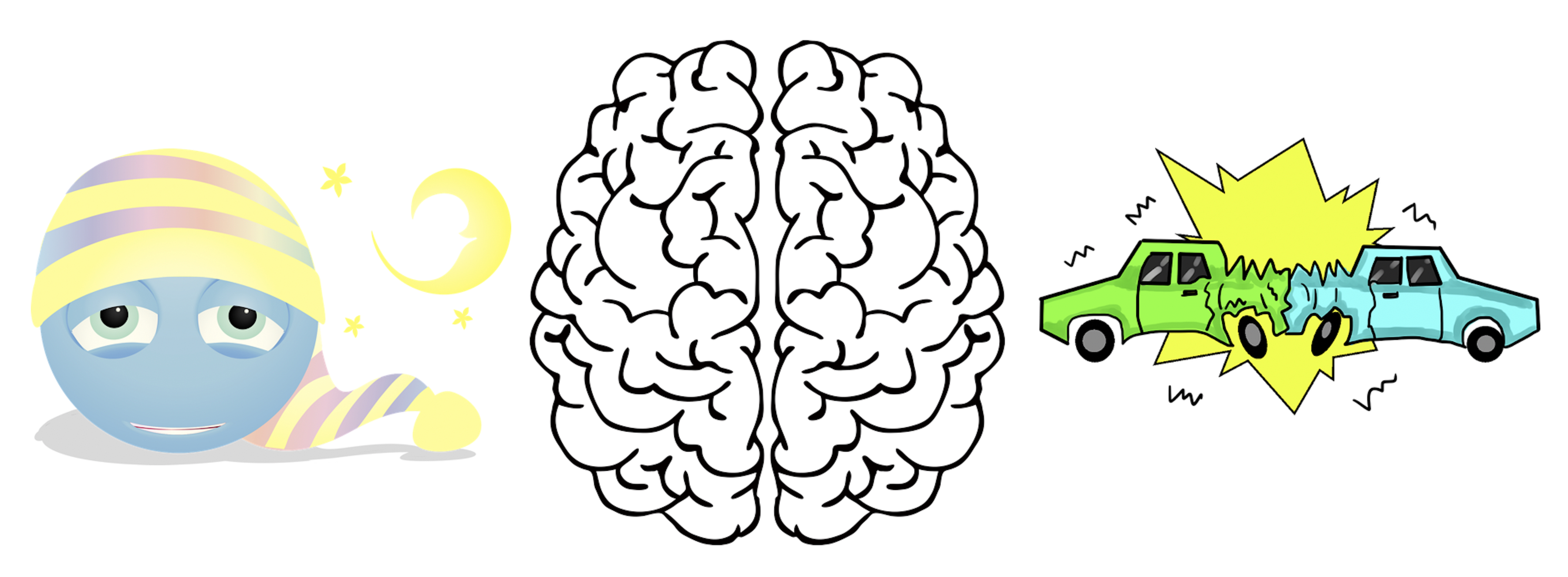
    Psychomotor impairments related to sleep deprivation directly affect driving ability. Researchers have found that driving while sleep deprived is similar to driving while under the influence. In fact, it is statistically equivalent to driving with a blood alcohol content level of being legally intoxicated. Those who drive while sleep deprived and those who drive while drunk have trouble maintaining their place on the road. They tend to accidentally cross into other lanes and look away from the road more often. Not surprisingly, the injury rate of motor crashes related to drowsy driving and drunk driving is similar.

    As students, we are often warned about the dangers of drunk driving and distracting driving (e.g., texting, talking on the phone), but there is little attention given to the dangers of driving while sleep deprived. The evidence is clear in regards to the importance of obtaining enough sleep for general health and cognitive function. However, if you must miss sleep, please refrain from driving. Instead, call a friend or call another ride. Just as you wouldn’t drive while under the influence, don’t drive while sleep deprived.

    References

    Centers for Disease Control and Prevention. (2016). 1 in 3 adults don’t get enough sleep. Atlanta, GA: US Department of Health and Human Services. Retrieved from https://www.cdc.gov/media/releases/2016/p0215-enough-sleep.html

    Durmer, J. S., & Dinges, D. F. (2005). Neurocognitive Consequences of Sleep Deprivation. Seminars in Neurology, 25, 1996. https://doi.org/10.1055/s-2005-867080

    Hershner, S. D. & Chervin, R. D. (2015). Causes and consequences of sleepiness among college students. Nature and Science of Sleep, 2014(6), 73–84.

    Watson, N. F., Badr, M. S., Belenky, G., Bliwise, D. L., Buxton, O. M., & Buysse, D. (2015). Joint consensus statement of the American Academy of sleep medicine and sleep research society on the recommended amount of sleep for a healthy adult: methodology and discussion. Sleep, 38(6), 591–592.

    Wilckens, K. A., Woo, S. G., Kirk, A. R., Erickson, K. I., & Wheeler, M. E. (2014). Role of sleep continuity and total sleep time in executive function across the adult lifespan. Psychology and Aging, 29(3), 658–665. https://doi.org/10.1037/a0037234

  • If I only had a…spine?

    For as long as I can remember, I have been obsessed with how people move. Now, hear me out. Even simple movements are fascinating if you really think about it. Electrical signals from your brain and spinal cord communicating with hundreds of muscles, forcing them to work together in a perfectly balanced symphony of contractions. All to maneuver our unwieldy skeletons gracefully through space.

    Do me a favor and stand up.

    For most of us, this movement feels like one of the simplest things we can do.

    Now, look at your legs.

    There are over 50 muscles below your hips alone. Yet, all these muscles just contracted in expert harmony to use the precise amount of force needed to move your body against gravity, all while maintaining near perfect balance. Precisely how we can perform these seemingly simple yet crucial movements on a whim is an active and exciting area of research, leading us toward innovation in movement rehabilitation, robotics, and beyond. These are movements we don’t even notice, like activating our muscles to breathe, blink, maintain our balance, or even walk. If you did notice these movements, you likely wouldn’t be able to focus on much else, transforming a simple grasp into an impossible and difficult task.

    Luckily, you don’t need your brain to do any of these things.

    More than just a cord

    Many people think of the spinal cord as just that, a cord. The cords and cables we typically interact with are charged with a very important but relatively simple task: bringing electricity from point a to point b. While the spinal cord is very important for bringing electrical signals from your brain to your muscles and organs, it does so much more.

    Picture an orchestra with a smart but rather lazy conductor performing for an audience. The musicians are like the motor neurons in the spinal cord, connecting to and contracting the muscles when the neurons fire, allowing you to move. Complicated musical pieces require guidance by our lazy conductor, just like throwing a dart or grasping an object requires guidance by the brain [1]–[4].  The audience’s cheers allow the orchestra to adapt, just like you use sensations from your body to improve or guide movements [5], [6].

    Yet, just like our experienced musicians don’t need the conductor to play simple or repetitive musical pieces, you don’t need your brain to perform “classic” movements. “The spinal cord is able to achieve so many behaviors by itself, completely isolated from the brain,” explains Dr. Cope, spinal cord neurobiology researcher and Georgia Institute of Technology professor.  “You can completely isolate the spinal cord in a living animal from the brain and it can walk on a treadmill [7], [8]. It can change speeds as the treadmill changes speed. You put an obstacle in its way, it can learn to lift its leg over that obstacle,” all without our lazy conductor [9].

    It was discovered in the early 1900’s that your motor neurons are fully capable of running the show [10]. “What that tells you is that there is this rich circuitry that in fact the whole motor system relies upon,” Dr. Cope explained. All in all, it looks like the spinal cord has the classics all worked out for you. Feel free to tell your conductor they can take the day off.

    How to run around like a chicken with its head cut off

    Unsurprisingly, experienced musicians are able to play complicated, intricate music without their conductor. Surprisingly to many, however, your spinal cord is able perform complicated, intricate behaviors without any input from the brain. How exactly are these behaviors possible? It’s all in the organization.

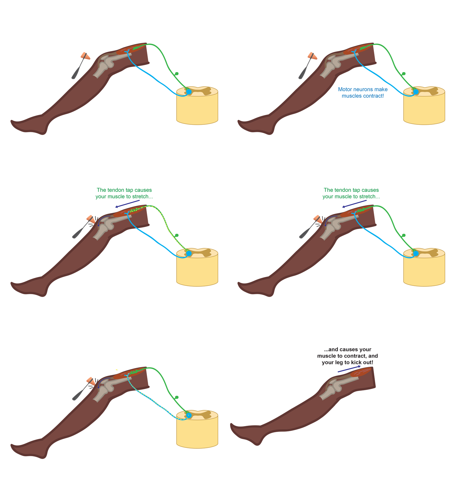
    If you’ve ever gotten a physical, you have probably experienced the odd sensation of your leg flying through the air without your consent. In the right spot, a simple tap on your knee by the doctor sends your foot on a trip automatically. We commonly refer to these types of movements as “reflexes,” in which no approval by the brain is required. This reflex pathway is relatively simple in organization; only two neurons are required, making this one of the fastest reflexes we have. One neuron senses the stretch of your muscle caused by the tap and immediately tells the second neuron to flex that same muscle. This flexion rapidly moves your leg before you can stop to think about it. This can happen in less than a few milliseconds [11]! You use your stretch reflex more often than your visits to the doctor, however. The stretch reflex helps you keep your balance without a second thought and is believed to be crucial for general sensory feedback and movement control.


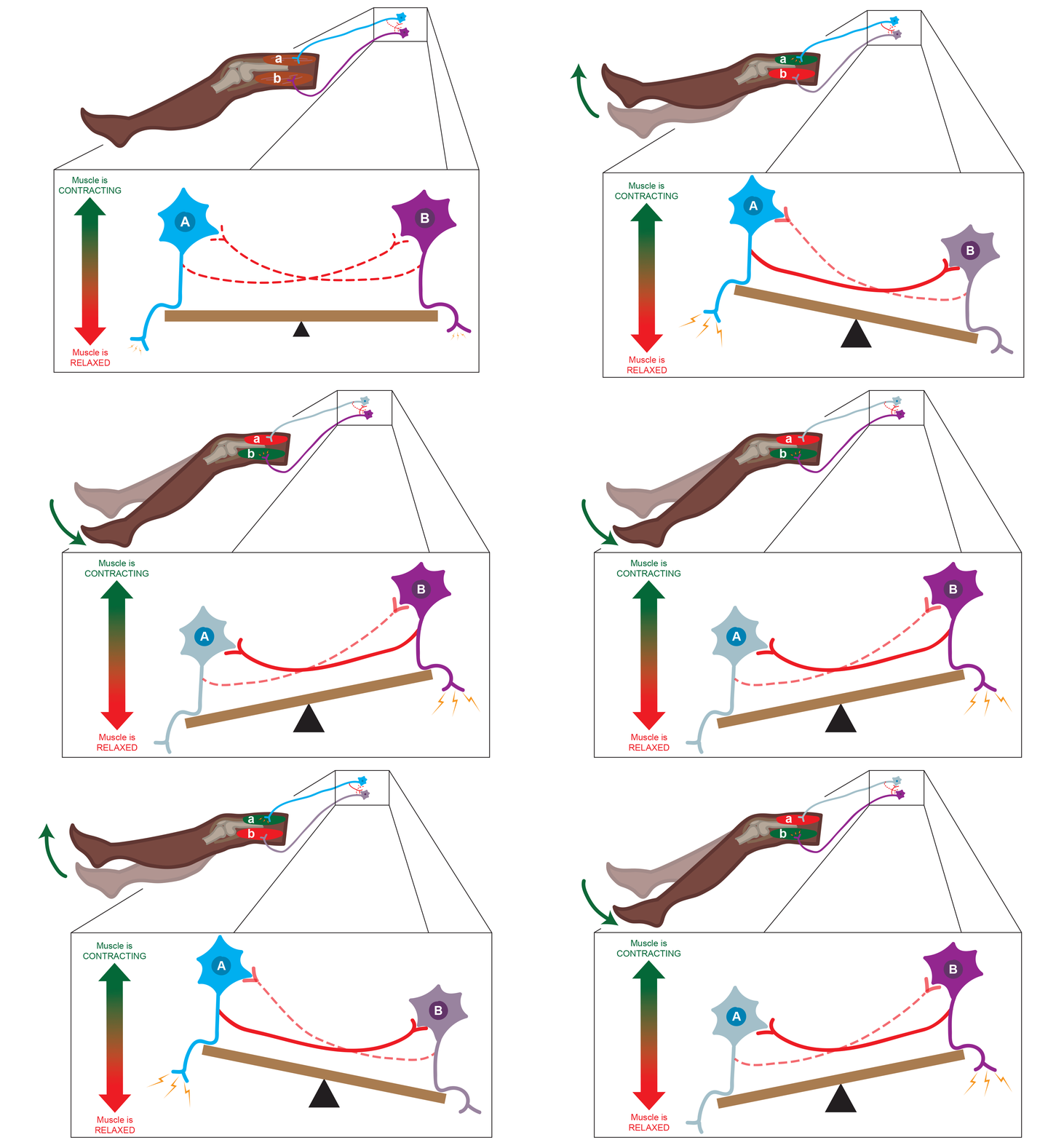
    What if we make things a little more complicated? Instead of just two neurons, let’s add in two sets of neurons in the spinal cord: set A controlling muscle a, and set B controlling muscle b. Much like a seesaw, these sets of neurons rhythmically alternate in activity. A neurons fire until they run out of juice, then B neurons take over and the cycle continues. With this small set of neurons, a pattern of alternating activity emerges. Together, A and B neurons form a central pattern generator. For humans, however, a 2-muscle central pattern generator isn’t very useful. Adding in more sets of neurons allows your spinal cord to rhythmically control more muscles in a more complicated pattern. With anything from breathing and scratching an itch to walking and running, the spinal cord is in charge.


    The patterns are there in your spinal cord, all you need to do is press start. “One of the things the brain does and can take full advantage of is to just send a ‘go’ signal to the spinal cord,” Dr. Cope explained. “[The brain] can say ‘Hey, all of the complicated things you do with timing and organizing … different muscles in different patterns, you do it. You’ve worked all that out. I don’t have to complicate my life with that.’” And while the brain can initiate and influence this pattern of alternating activity, it isn’t required. This pattern can just as easily be started by sensory input from your environment, or by sensory signals from throughout your body [9].

    At the end of the day, it seems like the spinal cord has it all figured out for us. But do we have the spinal cord all figured out? Not even close.

    The Mysterious Cord

    In the past year, the news has been abuzz with instances of paralyzed patients regaining the ability to walk [12]. Paralysis is typically caused by spinal cord damage. Up until recently it seems, spinal cord injuries often left patients with limbs that were difficult or impossible to move willingly, oftentimes without hope for improvement. So how are these patients taking these miraculous steps?

    A better question might be what happens to the spinal cord when it’s injured? We know some things about how it repairs itself, but we are far from the whole story. This means we are a far cry from fully repairing spinal cords ourselves. While these recent miraculous findings may make it seem like we have it all figured out, don’t let that fool you. “I think it’s exciting and I think it’s encouraging. I would say that we shouldn’t let our encouragement overshadow the fact that it’s nowhere close to what we want,” laments Dr. Cope.  “It’s going to require some basic neuroscience information about what the mechanisms are that are limiting recovery.”

    Researchers like Dr. Cope at Georgia Tech are working on a piece of this puzzle, studying to understand how the healthy and injured spinal cord contributes to and controls movement. Even with the great strides achieved recently by clinical studies [12], Dr. Cope explains that “We’re encouraged, but we have a long way to go.”

    References

    [1] D. P. Hanes and J. D. Schall, “Neural Control of Voluntary Movement Initiation,” Science (80-. )., vol. 274, no. 5286, pp. 427–430, 1996.

    [2] D. M. Wolpert and Z. Ghahramani, “Computational principles of movement neuroscience,” vol. 3, no. november, 2000.

    [3] D. A. Rosenbaum, K. M. Chapman, M. Weigelt, D. J. Weiss, and R. van der Wel, “Cognition, Action, and Object Manipulation,” Psychol. Bull., vol. 138, no. 5, pp. 924–946, 2013.

    [4] E. Freud, S. N. Macdonald, J. Chen, D. J. Quinlan, M. A. Goodale, and J. C. Culham, “Getting a grip on reality: Grasping movements directed to real objects and images rely on dissociable neural representations,” Cortex, vol. 98, pp. 34–48, 2018.

    [5] R. Shadmehr, M. A. Smith, and J. W. Krakauer, “Error Correction, Sensory Prediction, and Adaptation in Motor Control,” Annu. Rev. Neurosci., vol. 33, pp. 89–108, 2010.

    [6] D. M. Wolpert, Z. Ghahramani, and M. I. Jordan, “An internal model for sensorimotor integration,” Science (80-. )., vol. 269, no. 5232, pp. 1880–1882, 1995.

    [7] T. G. Brown, “The Intrinsic Factors in the Act o f Progression in the Mammal,” Proc. R. Soc. London. Ser. B, Contain. Pap. a Biol. character, vol. 84, no. 572, pp. 308–319, 1911.

    [8] S. Grillner and P. Zangger, “On the Central Generation of Locomotion in the Low Spinal Cat,” Exp. Brain Res., vol. 261, no. 34, pp. 241–261, 1979.

    [9] E. P. Zehr, “Neural Control of Rhythmic Human Movement : The Common Core Hypothesis,” Exerc. Sport. Sci. Rev., vol. 33, no. 1, pp. 54–60, 2005.

    [10] C. S. Sherrington, “Flexion‐reflex of the limb, crossed extension‐reflex, and reflex stepping and standing,” J. Physiol., vol. 40, no. 1–2, pp. 28–121, 1910.

    [11]  E. G. T. Liddell and C. S. Sherrington, “Reflexes in response to stretch (myotatic reflexes),” Proc. R. Soc. London. Ser. B, Contain. Pap. a Biol. Character, vol. 96, no. 675, pp. 212–242, 1924.

    [12] P. J. Grahn et al., “Enabling Task-Specific Volitional Motor Functions via Spinal Cord Neuromodulation in a Human With Paraplegia,” Mayo Clin. Proc., vol. 92, no. 4, pp. 544–554, 2017.

  • How The Science of Neuroplasticity Can Help You

    What is Neuroplasticity?

    Neuroplasticity seems like something a person would read about in a science-fiction novel. How can the brain, a seemingly unchanging mass of brain-goo, be constantly re-modelling and changing itself? As crazy as it seems, research has shown that throughout a person’s life, even as a person reaches adulthood, there are parts of the brain that remain “plastic” or moldable. For example, if a person loses their sight, the neurons associated with sight are re-wired to be used for other senses, such as touch or hearing. Although a child’s brain exhibits more neuroplasticity, the adult brain is still surprisingly flexible. This is specifically relevant to students, who use the brain’s natural ability to change for their own benefit.

    The Student Guide to Revamping Their Brain

    Feel less stressed, get better sleep, and feel more in control of your life!

    1.Meditation

    For most of neuroscience’s history, scientists believed that the brain was fixed or set. However, we now know that the brain changes itself in response to experiences, stimuli, thoughts, and the environment. Rather than being a static machine, the brain is constantly processing information and making new neural connections and letting old ones die. Evidence for this phenomena comes from the effect of meditation on the brain. Richard Davidson, a researcher from the University of Wisconsin, studied the effects of both short and long-term meditation practices. The researcher found that the meditator brain’s were different from the non-meditator brain’s. Not only did individuals who meditate feel less stressed, depressed, and more content, their actual brain showed differences too! On average, those who had a consistent mediation practice had more gray matter, which has been linked to mood control and decision making.

    2. Exercise

    If you need another reason to exercise, look so further! Exercise has been shown to not only increase spatial memory, but it promotes the generation and growth of neurons. People that exercise consistently have been shown to have a higher grey matter mass, especially grey matter associated with cognitive control. So, if you exercise, you might think and function better!

    3. Change Your Thoughts

    Although it’s surprising to think about, but the way we think can have an effect on our physical brain. When you think a thought, the brain forms a neural pathway. When you think that thought again, the neural pathway grows stronger. It is similar to the way a canyon is formed. Water flows through a canyon for years, making the path deeper and wider. Thought is powerful. When a person believes something, it has a physical manifestation in the brain. If a student believes that they can never be good at math, they will form a mental block. However, if they practice doing math, their brain will form a mini “map” for math and they will get better at it. The way we think can shape our worldview, what we do, and how we behave. It is good to be aware of that, even though a large part of our thoughts are not in our conscious awareness.

    Citations:

    Doidge, N. (2017). The brain that changes itself: Stories of personal triumph from the frontiers of brain science. Strawberry Hills, NSW: ReadHowYouWant.

    Lutz, A., Greischar, L. L., Rawlings, N. B., Ricard, M., & Davidson, R. J. (2004). Long-term meditators self-induce high-amplitude gamma synchrony during mental practice. Proceedings of the National Academy of Sciences, 101(46), 16369-16373. doi:10.1073/pnas.0407401101

    Begley, S. (2007, January 19). How Thinking Can Change the Brain. Retrieved from https://www.wsj.com/articles/SB116915058061980596


  • Badass Biology: Bat Echolocation

    Title

    Vision is awesomely complicated. A lot of animals, like the hawk, use vision to detect important environmental cues from vast distances. They use complex eyes and even more complex brains to locate prey, or figure out where the ground starts, or I dunno, check out lady hawks. Hearing is like vision’s less-popular but talented younger brother, the often-forgotten Luigi to evolution’s Mario. Hearing is amazing, yes, but our hearing kinda sucks. We’re not good at it, and so we discount its importance. Instead, we glorify vision as the pinnacle of evolutionary achievement, the ultimate sensory weapon against a hostile world. Yes, vision is awesome, and it is reliable, and it is useful for things like looking at steak. I f**king love steak. But other animals use hearing to see their steaks oh man that sounded really stupid so now it’s a run-on sentence now I don’t know how to end it oh no I’m embarrassed lol. The bat is one of the few animals that can accomplish this awkwardly-written steak hearing, and it does so using methods that absolutely amaze me.

    It must not come to a shock to you that bats use echoes from their own calls to create a mental picture of the world around them. Bats hear with the precision of a rocket-tipped arrow, which sounds totally stupid because that implies the rocket should blow the arrow backwards and that’s not where the pointy end is, but this is not important. I will explain to you how they use variable auditory frequencies, interaural timing, Doppler shift, and badass brain parts to make a map of their world. In short, they use magic. Bottom line, bats are badass.

    PARTS OF THE ECOCHOLOCATION PROCESS THAT ARE BALLS-OUT INSANE BECAUSE THEY GIVE BATS THE HEARING PRECISION OF A ROCKET-TIPPED ARROW.

    1-The call

    Did you also know that bats are really damn good at determining object size, velocity, shape, and even if it’s fluttering? I bet you didn’t, Mr. Fact Learner. You need to learn up all of your facts except that fact because you know for a fact that this fact is a fact, and that’s a fact.

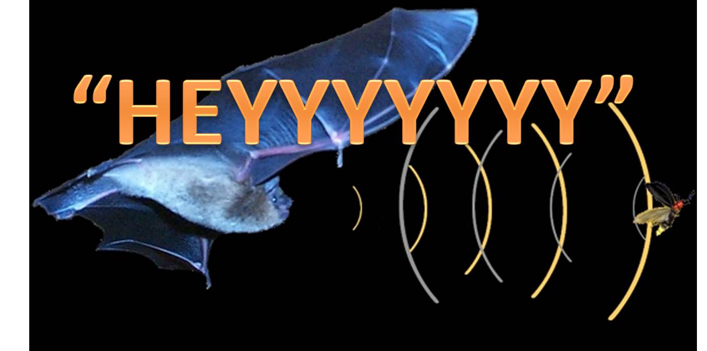
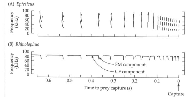
    Small bats usually emit two types of calls. First up are frequency-modulated (FM) sweeps, and bats use these to locate objects in the space around them. These calls are usually quick, and the pitch rapidly drops. This allows bats to emit a range of frequencies to the nearby area. The amount of time an FM sweep takes to return to the bat allows the bat to determine distance. Bats also use the time disparity between frequencies in the left and right ears to tell where objects are on the azimuth, or how left/right they are relative to the head.  The pointy shape of their ear also allows bats to use FM pulses to gauge the elevation of objects.

    We got three properties down in one paragraph: azimuth, elevation, and distance. Isn’t that enough? Hells nah. They use some of them AT THE SAME TIME. For instance, bats can tell the size of an object by determining how loud the echo is. They can then use all of this information to tell the difference between something like a bird that’s far away and a moth that’s up close. FM calls are sweet.

    The second type of call allows bats to tell how fast an object is moving. Constant-frequency (CF) pulses are typically longer, though the pitch does not drop. This allows, and yes I’m serious, bats to determine slight deviations in pitch due to the Doppler effect; a CF echo from an approaching object is slightly higher in pitch. This raises a good question: How can a bat do this if it’s flying toward an object? Wouldn’t the act of approaching an object make the CF pitch higher simply because the bat is moving towards it?

    FM and CF calls from two species of bats. Some species have FM and CF components within one call. Pretty sweet.
    FM and CF calls from two species of bats. Notice how quickly the pitch decays in the FM calls, whereas the CF calls sustain one pitch for some time. Some species even have FM and CF components within one call. Pretty sweet.

    This next experiment sounds kind of insane, but I promise it really happened.

    Scientists strapped a bat to a swing, pointed the swing at a wall, hooked up some microphones, and let loose. Sure enough, a bat swinging at a wall lowers the pitch of its emitted call. So, bats can compensate for their own movement through space by dropping the pitch of their call, making the pitch of the echo ALREADY ACCOUNT FOR DOPPLER SHIFT BY THE TIME IT GETS BACK TO THE BAT. Totally. Friggin. Badass.

    2-The ear and auditory pathway

    “Auditory pathway? Ughsheesh. That sounds boring. I don’t want to read about that.” said nobody awesome ever.

    Our inner ears are pretty complicated. Without getting stained with details, sound transduction works basically like this:

    1) Sounds, which are essentially air pressure waves, vibrate tiny bones in the middle ear that then convert those vibrations into pressure for the inner ear.

    2) The basilar membrane, a bendy flap of tissue in the cochlea that looks like an industrial-strength file, vibrates in different spots based on the frequency of these vibrations.

    3) The bending basilar membrane causes tiny hair cells adjacent to the cochlea to bend. These hair cells are connected to neurons that can detect when these hair cells bend.

    What did we learn here? Basically, pitch matters. Low pitches vibrate different hair cells than high ones. Bats are crazy though. Really crazy. I once saw one bite the head off a human.

    Bat basilar membranes, the part of the ear that transduces sounds into nerve impulses, are wider on the part of the cochlea where they detect the frequencies of their echoes. This helps for a number of reasons: 1) It increases the volume of noise in this frequency range, 2) It allows bats to become more sensitive to their own echoes, and 3) it helps reduce sensitivity to their own calls, reducing the odds of confusing those calls (or other sounds) with echoes. It’s also pretty cool to point out that bats contract muscles in their ears when they screech, dampening the sound of their calls further. All in all, bats are pretty much deaf to their own calls but highly sensitive to their echoes. Also, different bats are sensitive to different frequencies, limiting the odds of cross-talk between species.


    Left: Bats are sensitive to different frequencies. More hair cells at certain spots along the basilar membrane increase sensitivity. Right: The bat inner ear and auditory pathway.
    Left: Bats are sensitive to different frequencies. More hair cells at certain spots along the basilar membrane increase sensitivity. Right: The bat inner ear and auditory pathway.

    Alright, this is about to get nuts. I recommend not reading this if you’ve just had a heavy meal, because you may be so amazed that you’ll spew half-digested mush all over your keyboard.

    3- The brain

    I’m so excited to tell you about bat brains that I’m literally going to get some Oreos. It turns out I was actually excited about the Oreos all along.

    The auditory cortex is the part of the brain where auditory information is processed. You and I both have auditory cortexes. This is where information from the ears is organized and sent to other parts of the brain to aid in functions like cerebral processing, speech, and motor function. It’s also the part of the brain that allows us to differentiate between a high E flat played by a piano and a high E flat played by a chainsaw.

    Bats have auditory cortexes too. Theirs do many of the same things as ours. But there’s got to be a reason I’ve decided to spend an entire section on their brains. Trust me, there is.

    Remember how the cochlea can organize sounds by frequency? We tend to keep nerve impulses of similar frequencies bundled together as we send sound information from our ears to the auditory cortex through various brain structures. So, different parts of the auditory cortex become active when we hear different pitches, giving us somewhat of a tonal map of our sounds. Bats do this too, as do many animals.  Here’s the difference: bat brains are capable of producing an ACTUAL map based on sounds.

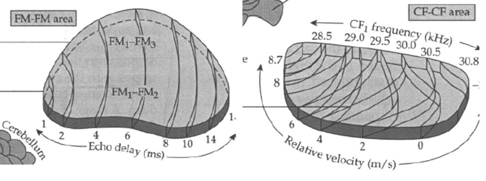
    The bat auditory cortex has two pretty badass regions. One is the FM-FM region. Remember FM pulses? Those are the calls bats use to determine where things are in the space around them. This function is done here. Neurons in the FM-FM area respond to the amount of time in the delay of a call and the harmonics of the call’s echo? What is a harmonic? Click here to find out.

    Think of the FM-FM area of the auditory cortex like a 2-D map. Each harmonic excites different horizontal sections from top to bottom, while the left-right axis responds to different delays. So, if the bat detects an echo from the 2nd harmonic after 8ms, neurons in a specific area of the FM-FM area will start firing. Enough of these allow a bat to tell how the large size and distance of the object is.

    Bats auditory cortexes can also produce a mental map of sounds based on their velocity. Recall that bats use CF-CF calls to detect  the Doppler Shift of sounds and tell whether they’re moving. They process this information in the CF-CF area of the auditory cortex. Like the FM-FM area, certain cells become active when sound properties are just right. Bands of cells respond to certain CF frequencies, and other bands respond to the velocity, or Doppler shift, of the sounds encoded. What bats end up with is a map of awesome.

    Left: FM-FM area from the mustached bat Pteronotus. Right: CF-CF area.
    Left: FM-FM area from the auditory cortex of the mustached bat Pteronotus. Right: CF-CF area.

    So what’s the point? Oreos can get me really excited. And to be specific, I’m talking about the golden ones. For some reason the regular Oreos get stuck in my teeth and I can never seem to get them out. Not that regular Oreos aren’t good, I’m just a little bit more partial to the ones that don’t annoy me. And bat brains, those are cool too. I wonder how they make the crème on the inside of the cookie. DO they keep it in big vats? I wonder how much would I have to bribe a guard to swim in that vat for 20 minutes.

    Could you imagine if I ended the article that way? Hahahahaha. I am so funny. But seriously, I am actually going to end the article this way. You don’t get a summary of what I said earlier.

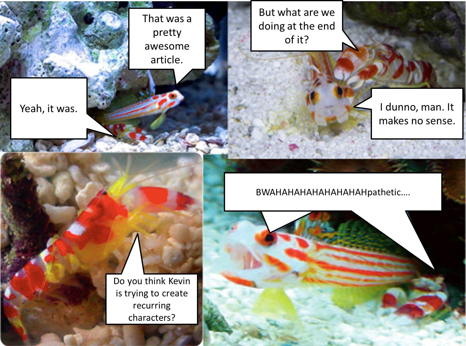
    Comic

    Want to suggest a topic for Badass Biology? Want to tell me how awesome a person you think I am? Interested in funding me? I didn’t think so. Anyway, leave a comment below and I might talk to you about stuff.

    A big shout-out to “Behavioral Neurobiology” by Thomas Carew for much of the information and figures used in this article. Dr. Carew, if you’re reading this, you seem like a cool guy. Email me and we will go grab a beer and some Oreos.


  • Neuroethics: expanding horizons

    I LOVE science. From genetics to astronomy, neuroscience to botany, this branch of knowledge endears me with its concrete facts, equations, and straightforward answers.

    Engineering? It’s cool too. As an engineering major in my second year, I’m in the process of learning a ton about what engineering actually is, and I’m enjoying it! You certainly have to know the science behind the technology you’re working on, but you also get to think creatively and innovate. On paper, you can have no limitations and let your designs go wild, but I’m learning that engineering in practice poses limitations that aren’t exactly fun to think about — cost and means of production for instance.

    Now, philosophy of science? That’s a whole new level for me. I’m currently taking a philosophy and public policy class on the topic of neuroethics. The problems we’re examining look at how new developments in science and technology can be implemented, with all the ethical, legal, and moral issues that come with it. These types of complex issues enter a completely grey area where there is no right answer. Until taking this class, I generally avoided these types of topics because they’re out of my comfort zone; I think of them as being “fuzzy,” whereas my beloved sciences are concrete.

    However, by taking this ethics class, I realized exploring these grey areas is essential. After all, if new discoveries in science are to come to any real world importance, they need to be applied in a practical, ethically sound way.

    Let me give you an example. The most recent problem my classmates and I tackled was that of using neuroimaging technology to identify students who are at risk for committing acts of violence. Specifically, we were targeting both bullying in schools and more serious matters such as school shootings.

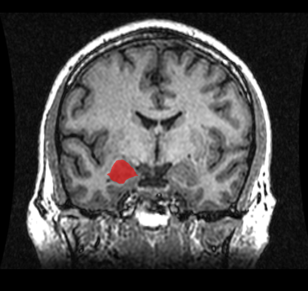
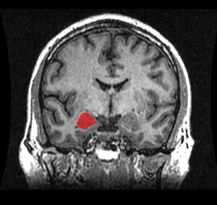
    MRI coronal view of the amygdala.
    Taken from the Wikimedia Commons.

    First, we examined the technology. (This part was really fun!) Recent neuroscience studies have examined the fMRI results of those who have committed various violent crimes (assault, homicide, etc.) and have been identified with various disorders (conduct disorder, antisocial personality disorder, etc.). Generally, results show the subjects to have decreased activity in certain regions of the brain as compared with the average person. These regions include the amygdala, prefrontal cortex, and the anterior cingulate cortex (ACC), all of which are part of the limbic system, or the “emotional brain.” (Sterzer, 2009). These brain abnormalities may be why some children, adolescents, and even adults do not empathize with others or even derive pleasure for putting others in pain.

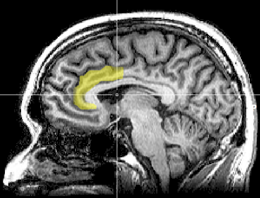
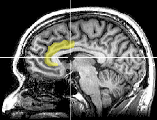
    Sagittal MRI slice with highlighting indicating location of the anterior cingulate cortex.

    So clearly there’s an inherent difference between the brains of violent and nonviolent individuals; shouldn’t we scan everyone for these defects and have them treated? Well not so fast. There is vast range of what “normal” is for a human brain. Scans have also found those with abnormal brain structure and no tendencies toward violence whatsoever. On the flip side of things, there are many violent criminals who lack these abnormal brain structures. If a scanning requirement were put in place for schools, false positives would create unnecessary stigma and hassle, and false negatives would allow students at risk for committing violence to slip through the system.

    On a related note, we need to consider that correlation does not equal causation. Just because a student may have a certain brain structure associated with violent tendencies, does not guarantee they will commit a violent act in the future. The brain is incredibly plastic, meaning it can change over time in response to experiences. An abnormal brain structure seen in a violent offender could be a result of actually committing the crime, or even a result of a negative childhood environment (Tsouderos, 2010).

    Beyond reading through the studies published on neuroimaging, we also had to look whether implementation of scanning in schools is financially, ethically, and legally sound. Have any of you ever had a PET, CAT, or MRI scan? Those things are expensive, especially if you don’t have health insurance. And in the case of a state program, that cost is placed on the tax payer. How accurate and effective do these scans have to justify paying thousands of dollars per scan? Once we have identified students as having an increased risk for violence, won’t we need the resources to treat them? And how do we manage the information taken from scans? Which school personnel, medical professionals, and authorities will have access to this information? Is it right to force a student to undergo a scan?

    In trying to find a solution, we ran into all of these questions and more. Ultimately, we decided against recommending using expensive neuroimaging technology (which, as cool as neuroscience is, it is still in its infancy) to combat violence in schools, instead looking at alternatives such as providing more resources for school programs to identify and treat mental disorders and promote mental health.

    However, whatever decisions we reached concerning this problem or the other topics in the class (ranging from genetic modification of humans to what type of medical information can be admitted into court), the important part of this experience was the discussion along the way. Thinking about these types of complex issues has really helped expand my thinking and round out my education. In fact, I was so consumed by our ethical debates that I’m seriously considering taking a proper introductory ethics or philosophy class. And to all you students out there, I recommend you do this as well! Or if not ethics, just something outside of your major and outside of your comfort zone; you never know when you’ll discover a new academic love!

    Sterzer, P., & Stadler, C. (2009) . Neuroimaging of aggressive and violent behaviour in children and adolescents. Frontiers in Behavioral Neuroscience, 3. Retrieved from http://www.ncbi.nlm.nih.gov/pmc/articles/pmc2766784/
    Tsouderos, T. (2010, Feb 15) . Exploring links between genes, violence, environment. Chicago Tribune. Retrieved from http://articles.chicagotribune.com/2010-02-25/news/ct-met-genetics-of-violence-20100225_1_genes-scientists-violence
    2. Taken from the Wikimedia Commons. Retrieved from http://upload.wikimedia.org/wikipedia/commons/1/10/MRI_anterior_cingulate.png
  • Facebook Friends: Why We Have So Many

    Facebook Friends: Why We Have So Many

    Me and my friends in a tour group in Amsterdam.

    You meet that hot rocker guy/girl at that concert you really like, and you guys trade names. You say, “Find me on Facebook”, and the next day or so, he/she adds you. You talk about wishing you were closer to the stage, the length of the concession lines, and why on Earth the T-shirt prices are so expensive. Typically, the conversation blossoms into a beautifully uninteresting synopsis of your daily routine: how tired you are despite normal hours of sleep, what YouTube links you think they’ll absolutely enjoy watching (because you do), and figuring out ways to make money without actually having a job.

    The chatter dwindles to a few comments or posts on one’s wall, then a “like”, and finally you’re down to that yearly “Happy Birthday (insert name here)” because you know they know its their birthday, and you don’t want to be that one guy/girl who doesn’t say anything. Why do we do this? Why don’t we talk to our friends anymore, and why have they forgotten us? Weren’t our YouTube links interesting?

    In the social media powerhouse we know as Facebook, I use the term “friend” lightly. Discussed by David McRaney in his blog, You Are Not So Smart, acquiring hundreds of friends on Facebook becomes a zero sum numbers game, with 150 as its approximate cap. You will typically be able to maintain a personal relationship with 150 people at any given time.

    The seemingly magical amount is the Dunbar number, developed by Robin Dunbar, a renowned anthropologist who correlated the size of the brain with the amount of potential social connections within a group. Even at this physiological limit, you’ll actively (on a daily or semi daily basis) talk to about 20. This doesn’t mean you’re a bad friend. You just have a limited neocortex.

    This cranial region takes into account social nuances and personal relationships. As your circle of friends increases, your maintenance of existing friendships increases several fold. Also, friends that have left your circle (be it geographic separation or interpersonal reasons) are harder to keep in contact with, as they require a portion of your processing power to keep the connection.

    The average person has 130 friends on Facebook (close to the Dunbar number), but there are people out there with +2,000. Do they talk to every single person they’ve ever friended? Of course not. It’s nearly impossible to do so with every other aspect of our lives screaming for attention. That’s not to say our social network isn’t a good thing. Facebook, Twitter, LinkedIn, and numerous other platforms for social networking are making communication easier than ever.

    We can have a great many loose connections to people, through our fascination with franchise coffeehouses, the latest Rube Goldberg contraption, or even that band that you like so much. 1 out of every 13 people on Earth is playing this never-ending game to increase their “friend stock”. Take solace in the fact that regardless of the number of Facebook friends we acquire, biologically, we’re basically average.