Origami does not originate from a sole inventor, it was crafted over centuries by many different hands for various ceremonial purposes in Japan and even China (Robinson, 2026). The word we use today, origami, is derived from the Japanese words ori which means folded and kami which translates to paper. While paper itself was first developed in China before spreading to Japan, folding paper into decorative and symbolic forms became especially refined in Japanese culture over time. Early uses of folded paper included religious ceremonies, gift wrapping, and formal exchanges, where presentation held great meaning.
Fast forwarding centuries, today origami is seen worldwide. A simple YouTube search nowadays can teach even the newest of the new how to craft an origami chameleon, like how I learnt! What was once a traditional practice has now grown into a global hobby, mathematical practice, and even an area of scientific research.
Math, Science, and Folds
It comes to no surprise when we see the intricate works by masterful artists, that the art within folding comes from mathematical roots. For example, take the common fold that basically all origami pieces start with: the square piece of paper folded diagonally corner to corner and then along the sides, top to bottom and left to right. These folds create lines of symmetry and reference points that help guide more intricate folds later on. Mathematically, these folds create either a valley or mountain based on the value of the angle between the two adjacent planes created by the fold (Kunz, 2024). A valley fold bends inward like a “V,” while a mountain fold rises outward like a mountain. By combining these two simple fold types in different orders, locations, and sizes, folders can create highly detailed animals, flowers, and geometric structures.
Additionally, origami also has strong roots in more of a geometric application of math, with many models and folds often relying on fractions, bisectors, symmetry and so much more. In the scientific world, origami has received its share of the spotlight from inspiring compact folded solar panel designs for satellite use to shaping medical stents used in patients.
My Folds and Why I do it
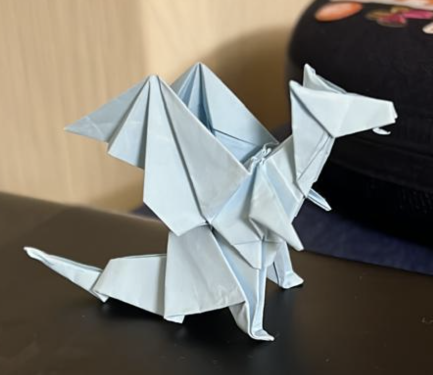
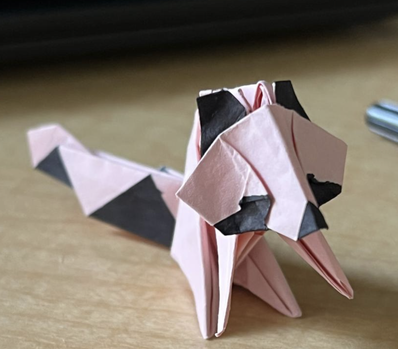
My goal when folding is simple: just don’t make it ugly. I find my inspiration, to recreate art through online tutorials, to be fueled by the passion and creativity of others. I folded the blue origami dragon below to surprise one friend, and the raccoon for another. I trace my folding skills back to one main motivation: wanting my creations to be as perfect as possible for the people around me. I have made plenty of lousy folds, but never when I feel a real sense of purpose behind them. When I fold for someone else, I tend to care more, focus more, and try as hard as I can. I am happy to say I will keep carrying on origami traditions. Mine may not be embedded in ceremonial, mathematical or even scientific purposes, but they still come from a place of creativity, curiosity, and humanity.
Clearly one article wasn’t enough to explore the full potential of organoids, so here we go again. This article won’t make much sense without reading the first but a quick catch up:
Organoids are 3D cell structures grown from Stem cell samples and grown in nutrient rich gel.
They can better model human systems as they are more complex than their 2D counterparts and a better representation of human systems than animal models.
This advantage makes organoids uniquely beneficial to the field of drug research and testing and personalized medicine.
Link to original article if desired: https://2026.chargedmag.org/2026/04/organoids-what-are-they-and-why-theyre-amazing/
Limitations
These applications are not without limitations however. While organoids can replicate many aspects of human tissue, some specific cell genes are harder to find in organoids (Andrews and Kriegstein, 2022). This impaired the organoids’ ability to properly simulate the systems they are derived from, preventing researchers from properly modeling diseases and treatments in that area.
Additionally, while developments have been made to improve organoid lifespans, organoids still require controlled environments and regular care to survive (Simian and Bissell, 2016). Even the longest lasting organoids, neurological models, have only survived a couple of years. Many organoids will even grow patches of dead tissue in the center due to a lack of oxygen hindering their research capabilities (Andrews & Kriegstein, 2022). In organic systems, cells are constantly interchanging molecules to maintain their functions and prevent necrosis. The nutritional gel can only do so much to get the molecules to cells at the interior of organoid structure without the natural systems to supply it.
All of this causes organoid maintenance to be costly compared to in-vitro models, which are relatively cheap and easy to handle (Schutgens & Clevers, 2020). This stifles research leading to many knowledge gaps in the field. Furthermore, these restrictions limit modeling larger systems. Potential treatments tested in smaller organoids could have unpredictable impacts on a larger system when prescribed. Concerns around this limits applications of organoids, creating Theory-Application Voids.
There are also many population gaps in organoid research as certain demographics are underrepresented in the stem cells used to create the organoids. These stem cells are often pulled from stem cell donor registries which have significant demographic, systemic, and behavioral biases. Further development into organoids and increased diversity in biobanks can increase organoid capabilities and applications.
Concerns
There are also current concerns surrounding organoid research that need to be addressed. While organoid models no longer rely on stem cells taken from embryos, they are still used in organoids modeling traditional embryo frameworks. These models use STEM cells to study the process of early embryonic development and can be very useful in identifying gestation concerns or potential risks to a fetus (Pereira et al., 2020). Once again, the line of when a zygote becomes a person however, gets blurry. Multiple public figures and many private citizens are debating if the creation of an embryo, even as a limited organoid, for study is ethical.
There are also concerns about neurological organoid models used for neurological diseases (Lavazza and Massimini, 2018). Current models can only imitate parts of the human brain. However, as models more closely resemble a human brain with increased functionality, ethical questions surrounding the creation and experimentation of something than can think and feel will only grow. The line being living, sentient, and sapient is hard to discern, especially with limited structures that might not have the ability to communicate mental facilities.
Future implementations of organoid models also raise ethical concerns regarding their financial exploitation and patient consent. People’s cell matter must be collected and added to databases to be used in organoid research (Bredenoord et al., 2017). Active patient involvement in organoid development is essential when considering this process. Their participation in cell donation must remain voluntary and properly compensated, and this is currently not guaranteed.
The implementation of organoids in personalized medicine could also have complications on the healthcare system (Stefanicka-Wojtas & Kurpas, 2023). While organoids could improve personalized medicine, the increased specificity of treatments could lead to decreased procedure lifespans and certainty. The underrepresentation and price of organoid backed research could also exacerbate existing disparities in healthcare and lead to an increase in general healthcare costs or dropped plans due to cost-shifting (What’s Behind Rising Health Insurance Costs? | Johns Hopkins Bloomberg School of Public Health, 2025).
Scientists in organoid research have repeatedly advocated for remaining cautious and careful in this relatively unexplored ethical field.
Potential Applications
Despite all of that dour news, organoids are still having a positive impact on millions of people with the potential to aid millions more. Unfortunately we only have time for two of these here: Regenerative Medicine and Transplantation.
Regenerative Medicine is the field of science dedicated to the regeneration and restoration of human tissue based in Stem cells. Many different animals have regenerative properties from the regrowth and axolotl limbs to sea cucumbers regrowing internal organs to flatworms regrowing entire heads. Throughout history scientists have been inspired by these properties and researched the mechanisms behind them, leading to the field of regenerative medicine. While the study of artificial organs has been around since the 1930s, the modern idea of regenerative medicine centered around Stem cells started in the 1990s (Sampogna et al., 2015).
As mentioned in my first article, the very purpose of Stem cells is to replace damaged, specialized cells. By comprehending the properties behind their functions, scientists can apply them to other areas. It is organoids that allow scientists to grow the cell cultures and tissue samples that are behind regenerative research. If someone has a large wound or damaged organ, an organoid of that structure could be applied and integrated into the person’s natural system. The organoid could be made from the person’s own cells, bypassing an immune response that comes with many non-biomaterials or transplants. This has already been proven promising. In one study, organoids made from the stem cells of mice suffering from inflammation and damage of their colon lining were successfully transferred onto the mice’s injured tissue. The cultured tissue covered the damage leading to a decrease in inflammation and harmful symptoms (Babak Arjmand et al., 2022). There are currently limitations, such as the delivery of nutrients mentioned above that directly challenge this field, but with more research scientists can find better ways for organoids to improve people’s lives through regenerative medicine.
Transplantation is a great example of the applications of regenerative medicine. As mentioned above, tissues and cell cultures can be grown using organoids. This is not the extent of their potential however. As organoid research develops, scientists are working to make larger and more complex models. Through assembloids, the combinations of different types of organoid (a topic for a later article), lab made organs or entire organ systems could be created from a single stem cell. In 2025, scientists were able to successfully transplant pig kidneys made from organoids (SMC Spain, 2025). In the future organoids could be used to transplant an entire human heart or cardiovascular system. An even farther yet still plausible possibility would be the creation of entire limbs for transplants with scientists already researching limb organoids (Tsutsumi & Mototsugu Eiraku, 2023). Approximately 5,600 people in the U.S. die each year simply waiting for a transplant in addition to the 30% of recipients who die within 10 years of a transplant due to complications such as transplant rejection (What Are the Complications of Transplantation?, 2019). Organoids have the potential to prevent these deaths, and while there are still complications due to the price and longevity of organoids, they will only improve with continued exploration.
Conclusion
Organoids are amazing. They have the ability to impact almost every field of biomedical science and save millions of lives. While this article does take a much more nuanced look at organoids, diving into their limitations and complications, scientists are aware of these issues and taking careful approaches to research. Improvements in organoids, especially to their limitations, can only be made if research is supported and continued. There is still so much potential for organoids that we have only just scratched the surface of and so many more applications that we haven’t even considered yet. Organoids have even been proven to work with many other biomedical instruments and tools, aiding in their development as well. The possibilities are endless.
Organoids are my passion, I’ve been interested in them for years now. Yet no one knew what I was going on and on about. I hope that these articles inspire curiosity and spread the wonders of organoids to others.
————————————————————————————————
Works Cited:
Title Image: Illustration created by Alexandra Marshall
Andrews, M. G., & Kriegstein, A. R. (2022). Challenges of Organoid Research. Annual Review of Neuroscience, 45(1), 23–39. https://doi.org/10.1146/annurev-neuro-111020-090812
Bredenoord, A. L., Clevers, H., & Knoblich, J. A. (2017). Human tissues in a dish: The research and ethical implications of organoid technology. Science, 355(6322). https://doi.org/10.1126/science.aaf9414
Lavazza, A., & Massimini, M. (2018). Cerebral organoids: ethical issues and consciousness assessment. Journal of Medical Ethics, 44(9), 606–610. https://doi.org/10.1136/medethics-2017-104555
Pereira, A. M., Popovic, M., Dondorp, W. J., Bustos, M. T., Bredenoord, A. L., Chuva, S. M., Susanne, Roelen, B. A. J., Guido, & Björn Heindryckx. (2020). Modelling human embryogenesis: embryo-like structures spark ethical and policy debate. Human Reproduction Update, 26(6), 779–798. https://doi.org/10.1093/humupd/dmaa027
Sampogna, G., Guraya, S. Y., & Forgione, A. (2015). Regenerative medicine: Historical roots and potential strategies in modern medicine. Journal of Microscopy and Ultrastructure, 3(3), 101–107. https://doi.org/10.1016/j.jmau.2015.05.002
Spain, S. (2025, October 31). For the first time, pig kidneys modified with human renal organoids are transplanted into pigs. SMC España. https://sciencemediacentre.es/en/first-time-pig-kidneys-modified-human-renal-organoids-are-transplanted-pigs#:~:text=Pig%20kidneys%20modified%20with%20human%20renal%20organoids
Schutgens, F., & Clevers, H. (2020). Human Organoids: Tools for Understanding Biology and Treating Diseases. Annual Review of Pathology: Mechanisms of Disease, 15(1), 211–234. https://doi.org/10.1146/annurev-pathmechdis-012419-032611
Simian, M., & Bissell, M. J. (2016). Organoids: A historical perspective of thinking in three dimensions. The Journal of Cell Biology, 216(1), 31–40. https://doi.org/10.1083/jcb.201610056
Stefanicka-Wojtas, D., & Kurpas, D. (2023). Personalised Medicine—Implementation to the Healthcare System in Europe (Focus Group Discussions). Journal of Personalized Medicine, 13(3), 380. https://doi.org/10.3390/jpm13030380
Tsutsumi, R., & Mototsugu Eiraku. (2023). How might we build limbs in vitro informed by the modular aspects and tissue-dependency in limb development? Frontiers in Cell and Developmental Biology, 11, 1135784–1135784. https://doi.org/10.3389/fcell.2023.1135784
What are the complications of transplantation? (2019, April). National Kidney Federation. https://www.kidney.org.uk/what-are-the-complications-of-transplantation#:~:text=Within%20one%20year%20of%20any,ten%20years%20after%20a%20transplant.
I’m willing to bet you’ve never heard of Organoids. No worries if so, they’re a pretty niche part of biomedical sciences. This is such a shame however as they’re having a great impact on research and our world. Organoids are often defined as tissue samples embedded into 3D extracellular matrix (ECM) gels or clonal derivatives of cells grown without mesenchyme and derived from stem cells. Don’t worry if your eyes have already glazed over! Mine did too the first time I read that.
In simpler terms, they are the growing of cells, taken from a patient, in a nutrient rich gel. While the field might seem niche at first, it has a long history and the potential to impact millions.
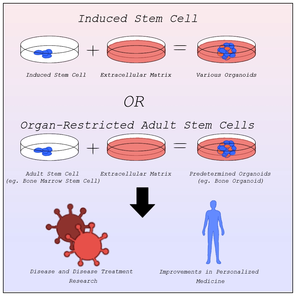
Before Organoids, most human physiological research relied on in vitro 2D cell culture and animal models (O’Connell & Winter, 2019). These often fail at clinical trials due to oversimplification of or incompatibility with human systems. At the time, cell cultures were limited to growing single layers of cells in a petri dish with none of the 3D complexities of real biological systems. This all changed with Ross Harrison’s “Hanging drop Cell Cultures” in 1906 (Simian and Bissell, 2016). By creating a 2D cell culture and turning the petri dish upside down, gravity would cause the cells to clump together and form a cohesive 3D spheroid of cells that could intermingle and interact like normal. Think about that! 3D structures could form by simply turning them upside down with no other interference. This was the start of research into organoids which offered a new way to look directly at human biology without risking patient or animal lives.
One key breakthrough was the discovery of 3D extracellular matrices first used in 1963 (Simian and Bissell, 2016). The gel provided a medium for natural scaffolding of cells rather than relying on gravity. This allowed for more complex 3D shapes and allowed for more types of organoids that more closely resembled complex real life systems. The gel also aided in providing the cells with nutrients required to keep them alive. Hence the “nutrient rich gel” from the definition above.
Exploration into this new field was rather slow however. The human body is composed of roughly 200 distinct types of cells, each of which specialize in a different function such as blood or nerve cells. Despite this, organoids mainly rely on only two types of cells, pluripotent stem cells and organ-restricted adult stem cells (Bredenoord et al., 2017). Stem cells are unspecified types of cells, yet to differentiate, with the ability to become any type of specified cell. They often serve to repair the human body as they can replace any damaged cells no matter the type. This ability makes them highly valuable to organoids trying to model systems that rely on the cooperation of multiple cell types.
Before the discovery of induced stem cells in 2006 by Dr. Shinya Yamanaka at Kyoto University, organoids were made from pluripotent stem cells as organ-restricted adult stem cells can be hard to access and are restricted to becoming cell types of their tissue of origin most commonly found in embryos (Agarwal et al., 2024). The line of when a zygote becomes a person, however, is blurry, raising many ethical concerns about the desecration of a person’s body. Induced stem cells allow for the change of specialized cells back into stem cells. Organoids can now be created from any tissue sample, entirely limiting the need for embryonic stem cells outside of studying early organ development (Frans Schutgens & Clevers, 2019).
With better ways to develop organoids, scientists quickly realized their potential. Organoids offer unparalleled advancement in disease research and treatment, personalized medicine, and so much more that we simply don’t have time for here.
Image by Alexandra Marshall
Disease Research and Treatment
One pivotal area of applications is understanding the development of diseases and disease treatment in humans. The lack of 3D structure in in vitro models lead to limited complexity and physiological relevance when studying diseases. Faulty predictions based on these studies can lead to treatments failing, with approximately 90% of clinical study drug candidates failing clinical trials and drug approval (Sun et al., 2022).
Organoid models have none of these limitations. In 2013, the first human condition modeled using organoids, cystic fibrosis, was published. The model had “excellent predictive value” and became the first successful personalized treatment method for cystic fibrosis patients (Lancaster and Huch, 2019). The modeling of other diseases using organoids quickly followed from genetic diseases like Polycystic kidney disease and Alagille syndrome, to infectious diseases like Covid-19 and the Zika virus, to neurological diseases like Alzheimer’s and Parkinson’s, to multitudes of cancers, and more (Xu et al., 2018). The ability for organoids to be made of any human system means they can model any disease regardless of which systems they afflict.
Organoids can be also grown co-culturally with bacteria allowing for bacterial infections and the diseases that arise from them to also be modeled and researched with organoids (Schutgens and Clevers, 2020). This has allowed for better understanding of bacteria like Helicobacter Pylori and Salmonella, aiding in detection and cures. As mentioned above, treatments can then be tested on these organoid systems without risking human lives or animal cruelty.
Respiratory Syncytial Virus (RSV), a disease that is being modeled in airway organoids, causes around 66,000–199,000 deaths among children annually (Schutgens & Clevers, 2020). This is just one of the many diseases for which scientists are now better equipped to find viable treatments. Organoids have already saved millions of lives and their potential for future disease treatment has the potential to millions more.
Personalized Medicine
While these treatments work for many people, genetic and biological differences can impact treatment effectiveness and tolerance with some even leading to negative side effects (Breen, 2021). In such situations, personalized medicine that is specialized to fit these differences might be necessary. Personalized medicine, sometimes called precision medicine, challenges the idea that treatments that pass general trials will work for everyone, instead working to construct a treatment plan with a specific patient in mind best suited to their physiology.
The idea of personalized medicine has been around for thousands of years, using one’s family’s history of diseases to diagnose the likelihood of that person having said disease. In the late 1900s the idea of evidence based medicine, the implementation of explicit reasoning based on a specific patient’s data, started being common practice when prescribing a patient (Visvikis-Siest et al., 2020). This opened the door for personalized medicine in modern science (Visvikis-Siest et al., 2020). This data was largely based on patients’ reactions to other treatments and family history however limited its effectiveness. The development of molecular biology and human genome mapping allows for a direct look at a patient’s physiology, aiding medical professionals in determining the biological reasons behind potential reactions to treatments before administration limiting adverse reactions.
Organoids have greatly furthered this field. As mentioned above, organoids are made from a patient’s induced stem cells. This means that the organoid culture into a structure based on the patient’s DNA, containing all the same biomarkers and physiology that the patient would have at that level. Where other treatments could have varying effects from the tested population, organoids allow medical professionals to see the exact effects a treatment plan would have on the patient. The use of stem cells also allows organoids to grow much quicker than other models. This allows for quick and accurate testing of the effects of medication on a patient. One study published in Science involving gastrointestinal cancer therapies found that organoids would correctly detect when drugs would fail 100% of the time, and when the drug would succeed 88% percent of the time (Vlachogiannis et al., 2018). Once again, because of the diversity of systems that organoids can model, organoids can be used to model many different personalized medicine plans allowing for the variety of treatment testing needed to find the most optimal plan.
Conclusion
Organoids have had a monumental, yet widely unknown, impact on a wide variety of disease advancement and treatment developments and personalized medicine. These structures can then be used to model human tissues and organs with much higher accuracy and complexity than previously used non-human or in vitro models.
Scientists use them to model disease development in humans and treatments designed for humans. This allows for a better understanding of the diseases and more effective treatments with fewer errors. They are also used to limit the risk of negative side effects from non-personalized medicine by furthering personalized medicine through the accuracy and speedy testing of many different treatment options.
With continued and careful development of organoids, disease understanding and treatments, personalized medicine and so much more can continue to be improved, saving millions of lives.
————————————————————————————————-
Work Cited:
Agarwal, S., Saha, L., Sohani, A., Karri, U., Gor, R., & Ramalingam, S. (2024). The importance of niche in therapeutic strategies targeting normal and cancer stem cells. Cancer Stem Cells and Signaling Pathways, 15–33. https://doi.org/10.1016/b978-0-443-13212-4.00031-3
Bredenoord, A. L., Clevers, H., & Knoblich, J. A. (2017). Human tissues in a dish: The research and ethical implications of organoid technology. Science, 355(6322). https://doi.org/10.1126/science.aaf9414
Frans Schutgens, & Clevers, H. (2019). Human Organoids: Tools for Understanding Biology and Treating Diseases. Annual Review of Pathology Mechanisms of Disease, 15(1), 211–234. https://doi.org/10.1146/annurev-pathmechdis-012419-032611
Generation and Cultivation of 3D Cell Models | ibidi. (2024). Ibidi. https://ibidi.com/content/1037-generation-and-cultivation-of-3d-cell-models
Lancaster, M. A., & Huch, M. (2019). Disease modelling in human organoids. Disease Models & Mechanisms, 12(7). https://doi.org/10.1242/dmm.039347
O’Connell, L., & Winter, D. C. (2019). Organoids: Past Learning and Future Directions. Stem Cells and Development, 29(5), 281–289.https://doi.org/10.1089/scd.2019.0227
Schutgens, F., & Clevers, H. (2020). Human Organoids: Tools for Understanding Biology and Treating Diseases. Annual Review of Pathology: Mechanisms of Disease, 15(1), 211–234. https://doi.org/10.1146/annurev-pathmechdis-012419-032611
Simian, M., & Bissell, M. J. (2016). Organoids: A historical perspective of thinking in three dimensions. The Journal of Cell Biology, 216(1), 31–40. https://doi.org/10.1083/jcb.201610056
Sun, D., Gao, W., Hu, H., & Zhou, S. (2022). Why 90% of clinical drug development fails and how to improve it? Acta Pharmaceutica Sinica B, 12(7), 3049–3062. https://doi.org/10.1016/j.apsb.2022.02.002
Visvikis-Siest, S., Theodoridou, D., Kontoe, M.-S., Kumar, S., & Marschler, M. (2020). Milestones in Personalized Medicine: From the Ancient Time to Nowadays—the Provocation of COVID-19. Frontiers in Genetics, 11. https://doi.org/10.3389/fgene.2020.569175
Vlachogiannis, G., Hedayat, S., Vatsiou, A., Jamin, Y., Fernández-Mateos, J., Khan, K., Lampis, A., Eason, K., Huntingford, I., Burke, R., Rata, M., Koh, D.-M., Tunariu, N., Collins, D., Hulkki-Wilson, S., Ragulan, C., Spiteri, I., Moorcraft, S. Y., Chau, I., & Rao, S. (2018). Patient-derived organoids model treatment response of metastatic gastrointestinal cancers. Science, 359(6378), 920–926. https://doi.org/10.1126/science.aao2774
Xu, H., Jiao, Y., Qin, S., Zhao, W., Chu, Q., & Wu, K. (2018). Organoid technology in disease modelling, drug development, personalized treatment and regeneration medicine. Experimental Hematology and Oncology, 7(1). https://doi.org/10.1186/s40164-018-0122-9
Have you ever seen something so cute you want to squeeze it? Maybe it was a puppy or kitten, or perhaps a teddy bear or doll. You may wonder why you feel this way – after all, it seems rather strange to mimic violence against something endearing. What makes cute things just so emotionally powerful, and what makes them cute in the first place?
Cuteness as Affect
In an article by the University of Oxford, Associate Professor Morten Kringelbach explains that cute things draw in our senses, attract our attention and spark rapid brain activity in the orbitofrontal cortex – a region associated with emotion in pleasure. In fact, the presence of something cute (in the case of this research article, a baby’s face) can activate the orbitofrontal cortex in 140 ms, or just under a seventh of a second (Kringelbach, 2016). Cuteness overloads the emotional state of the brain, drawing in mental attention and the five senses.
The feeling of cuteness, what researchers call its “affect,” has very interesting roots. From a biological perspective, an emotional response to cuteness may be derived from an evolutionary need to protect babies from harm. As Dr. Kringelbach explains, the evolved instinct to protect and care for babies may drive our wider perceptions of cuteness. Humans tend to express affection not only for babies and small children, but also for animals with baby-like features, and even dolls, teddy-bears, and miniature objects. These animals and objects trigger similar evolutionary protective desires – as well as a drawing in of attention and a general increase in emotional pleasure.
If cuteness is an evolutionary instinct to care and protect, why do we feel “cuteness aggression” – the “urge to squeeze or bite cute things” without causing harm (Alba and Stavropoulos, 2018)? Researchers at the University of California, Riverside, took on this challenge and made some interesting findings. Summarizing greatly, cuteness aggression was found to be an evolutionary response to the overwhelming emotions of experiencing cuteness (Alba and Stavropoulos, 2018). Evolutionarily, it would be less than ideal if early humans were “incapacitated” with emotion (as the researchers put it) in the presence of cuteness, and thus experienced a counter-emotion (scientifically called a “dimorphous expression” – when human action expresses the opposite feeling as to what they feel, like laughing when you cry). In the case of overwhelming cuteness, this dimorphous expression of playful harm held an evolutionary role to stop early humans from being too affected by cuteness.
Cuteness as Aesthetic
While this simple definition of cuteness – an evolutionary instinct to care for and be emotionally attached to babies (and by extension, baby-like animals and objects) – contains an incomplete and shallow understanding of how cuteness exists as a concept in society. As Joshua Paul Dale writes in his academic publication, The Aesthetics and Affects of Cuteness, “cuteness occupies both the aesthetic and affective realms” – and the modern extent of research places extensive focus on the affective side and avoids discussing the aesthetic. Note that in this usage, Dale does not mean aesthetic to mean cottage-core or punk – instead, the word is used within its sociological background, to understand how cuteness exists in the modern world.
Dale writes that the emotional response of cuteness is “manipulated for a variety of purposes” – including commercial, artistic, and social (Dale, 2016). The feeling of cuteness is emotionally powerful, and for that reason, is manipulated (malevolently and selfishly at times, yes, but also largely in acts of self-expression) to produce action. As Dr. Kringelbach discovered in her article for the University of Oxford, cuteness invites such powerful emotions that it drives the subject to action, causing them to put in work to continue to be able to receive and engage with the source (Kringelbach, 2016). Socially, cuteness has further effect than its core, biological and evolutionary response. In the modern world, cuteness causes the subject “catharsis” (as Dale writes) that fosters a need to protect a vulnerable object from harm (Dale, 2016). This effect of cuteness links it to vulnerability, a link that Dale dives into (in more depth than is relevant for this article) to understand the connections between violence and cuteness. Remember that cute thing you wanted to squeeze? The association between cuteness and vulnerability may be why. As Dale writes, cuteness is disarming and tender, but also “involves a cognitive reaction in which the power differential between subject and object comes into play” (Dale, 2016). Cuteness is, in many ways, an expression of a power dynamic.
So… what is cuteness? There’s the affective component: baby-like things trigger a biological response that draws attention and fosters feelings of care, and there’s the aesthetic component: cuteness is linked with vulnerability, intensity of emotion, and power dynamics, defining its socially constructed half. Together, these components create the feeling of cuteness that we understand today. It’s youthful, tender, disarming, attention-grabbing, and (in large part, but not entirely) biological.
References
Dale, J. P. (2016). The Appeal of the Cute Object: Desire, Domestication, and Agency. In The Aesthetics and Affects of Cuteness (1st ed., pp. 35–55). essay, Taylor & Francis Group.
Kringelbach, M. L. (2016). How cute things hijack our brains and drive behaviour University of Oxford. How cute things hijack our brains and drive behaviour. https://www.ox.ac.uk/research/how-cute-things-hijack-our-brains-and-drive-behaviour
Stavropoulos, K. K., & Alba, L. A. (2018). “It’s so cute I could crush it!”: Understanding neural mechanisms of cute aggression. Frontiers in Behavioral Neuroscience, 12. https://doi.org/10.3389/fnbeh.2018.00300
Since my first year at Georgia Tech, I’ve loved the campus’s architecture. There’s a perfect blend of old highlights that are the landmarks you think of when you think of GT, like Tech Tower and the Einstein statue, to newer buildings that represent the modern and futuristic side of campus, like the Kendeda Building for Innovative Sustainable Design and the renovated Price Gilbert Library and Crossland Tower.
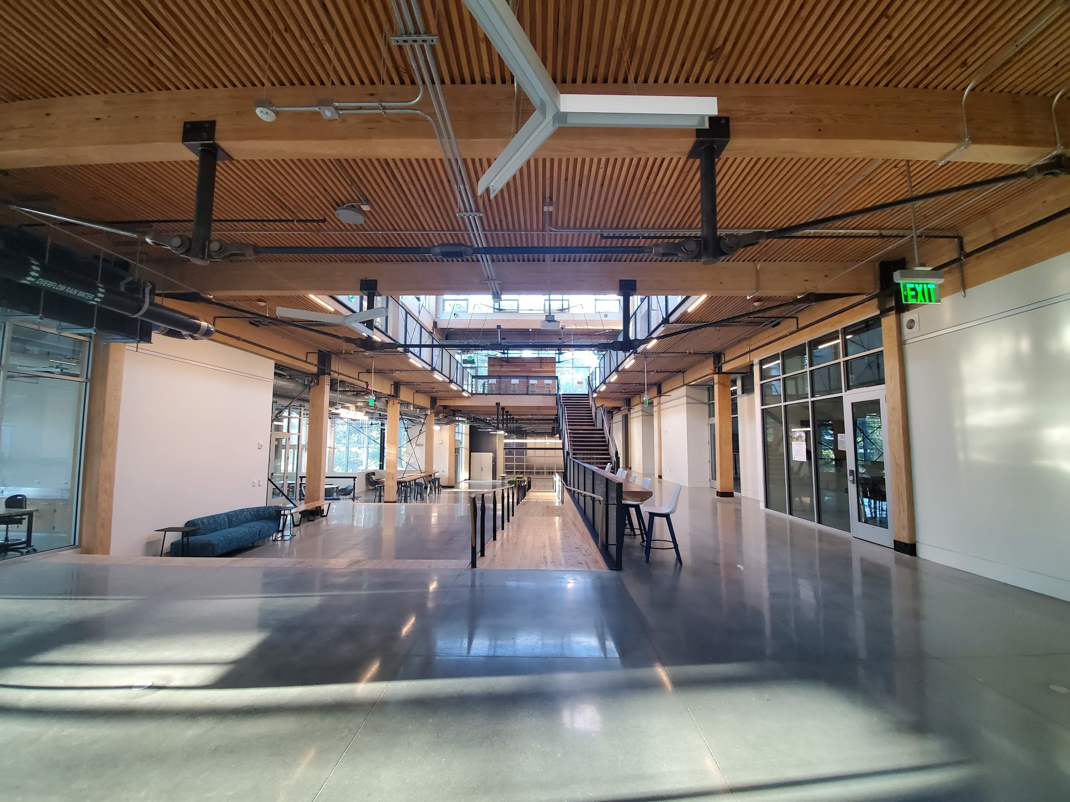
Inside the Kendeda Building for Innovative Sustainable Design.
When I lived in 8th Street Apartments last year, I noticed some exciting designs on each building’s entrance. Outside of 8th Street East, where I lived, there is a model of our solar system with the word “EXPLORATION” under it. I felt that the image was a beautiful tribute to the discovery of how the concept of our solar system developed since 1543. When I was walking near my old apartment a few weeks ago, I rediscovered that etching, and I wanted to take a deep dive into how many similar etchings were on campus. So with my friend, fellow Charged Magazine writer, and uber-talented photographer Steven Yang, we took a fun walk around West Campus, documenting everything we could find.
The Charged Dream Team, Steven Yang (back) and Jalen Borne (front) ready to explore GT’s campus.
During our journey, we found six different etchings at the entrances and the backs of student apartments on West Campus. Included were the solar system model mentioned earlier, the Georgia Tech seal, Leonardo da Vinci’s Vitruvian Man (with the word “HUMANITY”), the Earth representing “CIVILIZATION,” the Milky Way for “DISCOVERY,” and a beautiful but simple model of the atom, with “EXISTENCE.” At the Nelson-Shell apartment, there weren’t drawings, but the words “KNOWLEDGE” and “TECHNOLOGY” were marked. Finally, we found a plaque showing when Nelson-Shell was constructed for the 1996 Olympic Games.
What I loved about this adventure was discovering all of the etchings and gaining a deeper appreciation for what the pieces all together mean to me as a growing scientist. The etchings represent all the discoveries that have become the foundations of almost every STEM field and what they mean to society. Including knowledge and technology in the apartments perfectly complements the other buildings by considering the Georgia Tech motto: Progress and Service. We take the knowledge of the past, build new technology that improves the world, and further the values shown in the other campus etchings.
Campus etchings that Steven and I found throughout West Campus. Top row (left to right): Eighth Street East (back), Eighth Street South (front), Eighth Street South (back) Middle row: Nelson, Eighth Street West (back), Shell (back) Bottom row: Eighth Street East (back), Shell (front), Eighth Street West (front)
If you ever walk around campus and see a weird or cool sculpture or marking on the building, don’t just think about why it was added there, but what it means to you as a student. The etchings have become my favorite place to reflect on just how much campus has changed to become a place where its students work to bring the next generation of computer science, medical devices, and energy technology, all pushed by the motto of Progress and Service.
Born near Shanghai, China and later moving to the US, Chein-Shiung Wu was Chinese-American physicist at the forefront of nuclear physics and a contributor for the Manhattan Project. The bulk of her research surrounded that of beta decay, including publishing a still well regarded book on the topic in 1965. Wu was the first woman president of the American Physical Society in 1975 and the first woman recipient of an honorary doctorate from Princeton University.
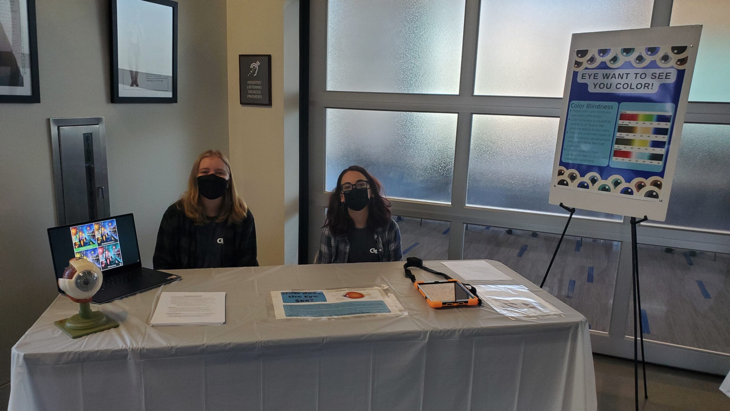
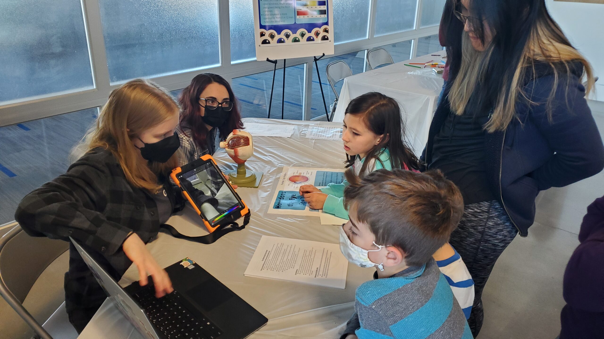
A couple of weeks ago our STEMComm Team put together an event for the Atlanta Science Festival called MindCraft. Here we presented several tables with fun arts and crafts that tied into different scientific concepts. My cohort had a table dedicated to the eye and how it views different colors. After going through the process of planning and setting up our station, there were many things I learned. Here are my top 5 tips to planning a science communications event:
Know your Audience
Being aware of who you will be presenting your content to is very important when considering what you will do. This involves asking yourself a series of questions such as, “How old will they be?”, “Are they new to these concepts?”, etc. By answering these questions, you will be able to cater your event to those specific needs and make it as effective as possible. For example, at our event, we knew that there would be some younger children in attendance so we made sure to have a station that they could more actively participate in by making our table’s craft a coloring sheet.
2. Find an Expert Collaborator
By finding someone that has more expertise in the area than you, you will be able to not only add to your content, but also get assistance in the overall set-up. At our particular station, we did not have a collaborator and did everything on our own which became a bit more stressful. We quickly learned from some of the other tables that did have collaborators that the work became much easier with more people to split the labor between. In order to find a good collaborator, there are again some questions you should ask yourself – “Who would have knowledge in my topic area?”, “What organizations/people do I have access to?”, etc. By reaching out to an organization that could assist you, you will have people that you can manage so that you get your event set up more efficiently and with less stress on yourself.
3. Create Eye-catching Visuals
Striking visuals are another important component of an event. This draws people in initially and gives them something interesting to look at while they take in your content. At our table, this became particularly important since we were talking about colors. With posters and flyers, we were able to display important diagrams and color scales so people could visualize what we were explaining. It is also important to interact with these visuals by pointing or gesturing towards them as it keeps your audience engaged in what you are presenting. In addition, it gives those waiting on your presentation something to look at while you speak to other guests.
4. Include Something Interactive
For an event such as these, interaction is key. By creating interaction between the audience and yourself as well as your content, people are more likely to learn something. For our particular table, we included an interactive coloring sheet that also included more information about the eye on the backside. This gave the guests something to physically do and apply the content to, plus, they were able to take it home with them! Having something to physically engage your audience with is important overall as it will keep them interested and help them apply and learn what you are presenting.
5. Rehearse Beforehand
When it comes to presenting information to others, it is important that you plan and rehearse what you will say in advance. If you do not do this, it is possible that you get stuck or do not present accurate information. In order to do this, you can set up some quick flashcards with information or use your presentation materials as guides. To rehearse, you can practice in the mirror, to your colleagues, or even to friends. This allows you to become comfortable with presenting the information and will make you sound more knowledgeable when you actually speak about it. However, you want to make sure that it does not sound like you are reading from a script as this could make your presentation boring. Rather, try to make it more casual and engage in conversations with your guests. Furthermore, you should be prepared for questions about your topic area. It is not the end of the world if you do not know how to answer something, but overall having done your research beforehand is important so that you are able to answer questions.
All in all, many components go into planning and setting up a successful science communications event. By following these 5 tips, you can ensure better preparedness and less stress while planning.
If you want to find out more about the specific materials and content we presented at our eyeball and color station, including tips on creating posters, flyers, and interactive worksheets, be on the lookout for another article coming soon! Best of luck with your event!
More Resources for Planning a Science Outreach Event:
You may have heard that modern birds evolved from dinosaurs before. More specifically, modern birds descend from theropods, which is a clade of dinosaurs. A clade is a group of species that share some key traits as well as common ancestry. Theropods all had hollow bones and three toed limbs, traits that remain in modern birds. The most famous of theropods was the T. Rex, but you also may have heard of the Spinosaurus, Archaeopteryx, and Allosaurus.
How did we come to this conclusion? It turns out hollow bones and three toed limbs are not the only similarities between theropods and modern birds. Various types of theropods have also been found to have feathers in fossilized remains. Older theropod species, that are “farther” back from dinosaurs on the tree of life, only had one kind of feather, which paleontologists think was used for insulation. Younger theropods, more closely related to birds, such as the oviraptors, had multiple different kinds of feathers. While some of these feathers are also for insulation, others are very similar to the flight feathers on modern birds. Modern birds have three “fingers” in the bone structure that supports their wings. Groups of dinosaurs that are theorized to be closely related to modern birds tend to have less and less fingers. The Orinsthican dinosaurs, very distant ancestors to birds, had five fingers. The Allosaurids, closer ancestors, had four fingers. Eventually, dinosaurs like Archaeopteryx had three fingers, which would eventually lead to the development of modern birds’ wings. Over time, the tails of dinosaurs began to get shorter and shorter. Eventually, the tails got so short that they’re barely there, until the only modern evidence of them are the tail feathers on the backs of birds.
References
Agnolin, F. L., Motta, M. J., Brissón Egli, F., Lo Coco, G., & Novas, F. E. (2019). Paravian phylogeny and the dinosaur-bird transition: An overview. Frontiers in Earth Science, 6(February), 1–28. https://doi.org/10.3389/feart.2018.00252
The authors of “Enriched environment exposure accelerates rodent driving skills”, published in Behavioral Brain Research, conducted an experiment intended to study the impact of the environment on the efficacy of stress-releasing mental health treatments in lab rats. This study aimed to see how an enriched environment, both socially and intellectually, promotes stress relief while doing fun activities. The process for teaching the rats to drive the rodent operated vehicles (known as ROVs) also has implications for the importance of enrichment in how service dogs are trained.
Study Summary
There were two groups of rats that participated in this study. The control group of rats lived in a standard environment for laboratory rats, in standard laboratory cages, each with one other rat. The experimental group of rats lived in housing with an enriched environment. These rats got multiple rat friends, rat toys to keep them occupied, and multi-level housing.
The study began with a “training period”, where rats in both groups were taught by operant conditioning how to drive the ROVs. While the authors of the study expected the rats living in standard housing to perform worse than the other rats, they were shocked at how much worse they performed. The authors noted that the control group rats failed to perform even “basic” tasks in driving the ROVs.
After the training period (in which food rewards were used to encourage the rats to drive), both groups of rats continued to drive the ROVs even in the absence of rewards. However, rats living in enriched housing drove the ROVs without treats for a longer period of time than the rats living in standard housing. Throughout the study, the scientists measured the levels of hormones that were indicative of the rats’ ability to handle stress. Both groups showed an improved ability to handle stress as a result of ROV driving, but the enriched housing rats showed a significantly larger benefit than the standard housed rats.
Conclusion
This investigation shows that the environment of an animal has a significant impact on the effect of stress relieving interventions. The authors suspect that the enriched rats were more able to learn new behaviors (driving the ROVs) as a result of experiencing a wider variety of behaviors in their enriched home environments. The results of this study have interesting ramifications on the optimal conditions for other animals who will be undergoing rigorous training. Additionally, this study demonstrates how a good environment that has opportunities for social connection and mental challenges is beneficial in mental health treatment.
References
Crawford, L. E., Knouse, L. E., Kent, M., Vavra, D., Harding, O., LeServe, D., Fox, N., Hu, X., Li, P., Glory, C., & Lambert, K. G. (2020). Enriched environment exposure accelerates rodent driving skills. Behavioural Brain Research, 378, 112309. https://doi.org/10.1016/j.bbr.2019.112309
Great blue herons are part of the Ardea genus, which is a genus of birds that includes the herons, egrets, and bitterns. These birds are typically pretty large in size, with lengths usually greater than 80–100 cm. You’re most likely to find members of this genus in wetlands and other partially aquatic habitats.
Feeding Behavior
Great blue herons are carnivores, and they eat a variety of aquatic, semi-aquatic, and terrestrial prey. Their diet consists of predominantly fish, but they will eat pretty much anything that they can catch in their beaks. Fish are supplemented by amphibians, reptiles, small ground mammals, and even other birds. Great blue herons catch smaller prey in the jaws at the back of their beak, although they prefer to stab larger prey with their beaks.
There are 38 total feeding behaviors that have been recorded by members of the Ardea family. Each species in this genus uses multiple feeding methods — they’re creative hunters. Twelve of these strategies have been observed in great blue herons. The most commonly observed feeding behavior for the great blue heron is standing and waiting, where the heron simply stands still and waits for its prey to come to it. Herons also will move slowly, either through the water or through a field, in search of prey. More dramatically, the great blue heron will occasionally plunge-dive, diving down quickly and with force, to catch its prey. A particularly interesting feeding behavior of the great blue heron is to stick its bill in the water, and vibrate it. The underwater vibrations create ripples, attracting fish and other aquatic prey.
Habitat
Great blue herons prefer to live in wetlands, especially when nesting. Rather than nesting alone, great blue herons nest in colonies. Colony nesting is particularly common for birds that live in aquatic environments. Colonies can have over 500 nests (so easily over a thousand birds) all relatively close together. For large scale habitats and especially for colony nesting, great blue herons have preferences for certain types of habitats over others. The lacustrine wetlands is a particular favorite of Illinois great blue herons. Lacustrine wetlands are any wetland or deepwater area that has these three characteristics:
Located in a valley/depression in the ground
Lacking trees, shrubs, and other plant life that covers more than 30% of the area
At least 20 acres in surface area
Biologists believe that herons may prefer this biome for their habitats due to the easy access to their preferred prey of small bodied vertebrates. Additionally, large wetlands tend to be isolated from human activity, which great blue herons tend to avoid if possible.
References
Combs, B. P. (2021). Great Blue Heron Forages for Fish by Bill-Vibrating. Western North American Naturalist, 81(2), 280–284. https://doi.org/10.3398/064.081.0213
Gibbs, J. P., & Kinkel, L. K. (1997). Determinants of the Size and Location of Great Blue Heron Colonies. Colonial Waterbirds, 20(1), 1–7. https://doi.org/10.2307/1521757
Knight, E. C., Vennesland, R. G., & Winchester, N. N. (2016). Importance of Proximity to Foraging Areas for the Pacific Great Blue Heron (Ardea herodias fannini) Nesting in a Developed Landscape. Waterbirds, 39(2), 165–174. https://doi.org/10.1675/063.039.0207