Have you ever wondered how scientists look at proteins or DNA? Here’s the secret- they don’t actually look at physical molecules all that often! Frequently, they use softwares like PyMOL. PyMOL is a widely used molecular visualization program that converts structural data into detailed 3D models, allowing scientists to rotate, zoom, and explore molecules interactively. It was originally developed by Warren Lyford DeLano and has become a standard tool in structural biology and chemistry (DeLano, 2002).
PyMOL works by reading files that contain the exact positions of atoms in three-dimensional space, often obtained from experimental techniques like X-ray crystallography and stored in databases such as the Protein Data Bank. Using this coordinate data, the program reconstructs molecules and displays them in different visual styles, including sticks, spheres, and ribbon diagrams that highlight protein folding patterns. These visualization approaches are essential for understanding how molecular structure relates to function and are widely used in both research and education (Sehnal et al., 2021).
What makes PyMOL especially powerful is how interactive and customizable it is. Scientists can highlight specific regions of a molecule, measure distances between atoms, and even model how molecules might interact with each other- something that is especially important in drug design and biochemistry. Because it translates complex data into intuitive visuals, PyMOL helps bridge the gap between abstract molecular concepts and real-world understanding, making it a valuable tool not just for scientists, but also for students learning about the molecular world.
References:
DeLano, W. L. (2002). PyMOL: An open-source molecular graphics tool. San Carlos, CA: DeLano Scientific. Available at http://www.pymol.org
Sehnal, D., Bittrich, S., Deshpande, M., Svobodová Vařeková, R., Berka, K., Bazgier, V., Velankar, S., Burley, S. K., Koča, J., & Rose, A. S. (2021). Mol* Viewer: Modern web app for 3D visualization and analysis of large biomolecular structures. Nucleic Acids Research, 49(W1), W431–W437. https://doi.org/10.1093/nar/gkab314
Berman, H. M., Westbrook, J., Feng, Z., Gilliland, G., Bhat, T. N., Weissig, H., Shindyalov, I. N., & Bourne, P. E. (2000). The Protein Data Bank. Nucleic Acids Research, 28(1), 235–242. https://doi.org/10.1093/nar/28.1.235
Clearly one article wasn’t enough to explore the full potential of organoids, so here we go again. This article won’t make much sense without reading the first but a quick catch up:
Organoids are 3D cell structures grown from Stem cell samples and grown in nutrient rich gel.
They can better model human systems as they are more complex than their 2D counterparts and a better representation of human systems than animal models.
This advantage makes organoids uniquely beneficial to the field of drug research and testing and personalized medicine.
Link to original article if desired: https://2026.chargedmag.org/2026/04/organoids-what-are-they-and-why-theyre-amazing/
Limitations
These applications are not without limitations however. While organoids can replicate many aspects of human tissue, some specific cell genes are harder to find in organoids (Andrews and Kriegstein, 2022). This impaired the organoids’ ability to properly simulate the systems they are derived from, preventing researchers from properly modeling diseases and treatments in that area.
Additionally, while developments have been made to improve organoid lifespans, organoids still require controlled environments and regular care to survive (Simian and Bissell, 2016). Even the longest lasting organoids, neurological models, have only survived a couple of years. Many organoids will even grow patches of dead tissue in the center due to a lack of oxygen hindering their research capabilities (Andrews & Kriegstein, 2022). In organic systems, cells are constantly interchanging molecules to maintain their functions and prevent necrosis. The nutritional gel can only do so much to get the molecules to cells at the interior of organoid structure without the natural systems to supply it.
All of this causes organoid maintenance to be costly compared to in-vitro models, which are relatively cheap and easy to handle (Schutgens & Clevers, 2020). This stifles research leading to many knowledge gaps in the field. Furthermore, these restrictions limit modeling larger systems. Potential treatments tested in smaller organoids could have unpredictable impacts on a larger system when prescribed. Concerns around this limits applications of organoids, creating Theory-Application Voids.
There are also many population gaps in organoid research as certain demographics are underrepresented in the stem cells used to create the organoids. These stem cells are often pulled from stem cell donor registries which have significant demographic, systemic, and behavioral biases. Further development into organoids and increased diversity in biobanks can increase organoid capabilities and applications.
Concerns
There are also current concerns surrounding organoid research that need to be addressed. While organoid models no longer rely on stem cells taken from embryos, they are still used in organoids modeling traditional embryo frameworks. These models use STEM cells to study the process of early embryonic development and can be very useful in identifying gestation concerns or potential risks to a fetus (Pereira et al., 2020). Once again, the line of when a zygote becomes a person however, gets blurry. Multiple public figures and many private citizens are debating if the creation of an embryo, even as a limited organoid, for study is ethical.
There are also concerns about neurological organoid models used for neurological diseases (Lavazza and Massimini, 2018). Current models can only imitate parts of the human brain. However, as models more closely resemble a human brain with increased functionality, ethical questions surrounding the creation and experimentation of something than can think and feel will only grow. The line being living, sentient, and sapient is hard to discern, especially with limited structures that might not have the ability to communicate mental facilities.
Future implementations of organoid models also raise ethical concerns regarding their financial exploitation and patient consent. People’s cell matter must be collected and added to databases to be used in organoid research (Bredenoord et al., 2017). Active patient involvement in organoid development is essential when considering this process. Their participation in cell donation must remain voluntary and properly compensated, and this is currently not guaranteed.
The implementation of organoids in personalized medicine could also have complications on the healthcare system (Stefanicka-Wojtas & Kurpas, 2023). While organoids could improve personalized medicine, the increased specificity of treatments could lead to decreased procedure lifespans and certainty. The underrepresentation and price of organoid backed research could also exacerbate existing disparities in healthcare and lead to an increase in general healthcare costs or dropped plans due to cost-shifting (What’s Behind Rising Health Insurance Costs? | Johns Hopkins Bloomberg School of Public Health, 2025).
Scientists in organoid research have repeatedly advocated for remaining cautious and careful in this relatively unexplored ethical field.
Potential Applications
Despite all of that dour news, organoids are still having a positive impact on millions of people with the potential to aid millions more. Unfortunately we only have time for two of these here: Regenerative Medicine and Transplantation.
Regenerative Medicine is the field of science dedicated to the regeneration and restoration of human tissue based in Stem cells. Many different animals have regenerative properties from the regrowth and axolotl limbs to sea cucumbers regrowing internal organs to flatworms regrowing entire heads. Throughout history scientists have been inspired by these properties and researched the mechanisms behind them, leading to the field of regenerative medicine. While the study of artificial organs has been around since the 1930s, the modern idea of regenerative medicine centered around Stem cells started in the 1990s (Sampogna et al., 2015).
As mentioned in my first article, the very purpose of Stem cells is to replace damaged, specialized cells. By comprehending the properties behind their functions, scientists can apply them to other areas. It is organoids that allow scientists to grow the cell cultures and tissue samples that are behind regenerative research. If someone has a large wound or damaged organ, an organoid of that structure could be applied and integrated into the person’s natural system. The organoid could be made from the person’s own cells, bypassing an immune response that comes with many non-biomaterials or transplants. This has already been proven promising. In one study, organoids made from the stem cells of mice suffering from inflammation and damage of their colon lining were successfully transferred onto the mice’s injured tissue. The cultured tissue covered the damage leading to a decrease in inflammation and harmful symptoms (Babak Arjmand et al., 2022). There are currently limitations, such as the delivery of nutrients mentioned above that directly challenge this field, but with more research scientists can find better ways for organoids to improve people’s lives through regenerative medicine.
Transplantation is a great example of the applications of regenerative medicine. As mentioned above, tissues and cell cultures can be grown using organoids. This is not the extent of their potential however. As organoid research develops, scientists are working to make larger and more complex models. Through assembloids, the combinations of different types of organoid (a topic for a later article), lab made organs or entire organ systems could be created from a single stem cell. In 2025, scientists were able to successfully transplant pig kidneys made from organoids (SMC Spain, 2025). In the future organoids could be used to transplant an entire human heart or cardiovascular system. An even farther yet still plausible possibility would be the creation of entire limbs for transplants with scientists already researching limb organoids (Tsutsumi & Mototsugu Eiraku, 2023). Approximately 5,600 people in the U.S. die each year simply waiting for a transplant in addition to the 30% of recipients who die within 10 years of a transplant due to complications such as transplant rejection (What Are the Complications of Transplantation?, 2019). Organoids have the potential to prevent these deaths, and while there are still complications due to the price and longevity of organoids, they will only improve with continued exploration.
Conclusion
Organoids are amazing. They have the ability to impact almost every field of biomedical science and save millions of lives. While this article does take a much more nuanced look at organoids, diving into their limitations and complications, scientists are aware of these issues and taking careful approaches to research. Improvements in organoids, especially to their limitations, can only be made if research is supported and continued. There is still so much potential for organoids that we have only just scratched the surface of and so many more applications that we haven’t even considered yet. Organoids have even been proven to work with many other biomedical instruments and tools, aiding in their development as well. The possibilities are endless.
Organoids are my passion, I’ve been interested in them for years now. Yet no one knew what I was going on and on about. I hope that these articles inspire curiosity and spread the wonders of organoids to others.
————————————————————————————————
Works Cited:
Title Image: Illustration created by Alexandra Marshall
Andrews, M. G., & Kriegstein, A. R. (2022). Challenges of Organoid Research. Annual Review of Neuroscience, 45(1), 23–39. https://doi.org/10.1146/annurev-neuro-111020-090812
Bredenoord, A. L., Clevers, H., & Knoblich, J. A. (2017). Human tissues in a dish: The research and ethical implications of organoid technology. Science, 355(6322). https://doi.org/10.1126/science.aaf9414
Lavazza, A., & Massimini, M. (2018). Cerebral organoids: ethical issues and consciousness assessment. Journal of Medical Ethics, 44(9), 606–610. https://doi.org/10.1136/medethics-2017-104555
Pereira, A. M., Popovic, M., Dondorp, W. J., Bustos, M. T., Bredenoord, A. L., Chuva, S. M., Susanne, Roelen, B. A. J., Guido, & Björn Heindryckx. (2020). Modelling human embryogenesis: embryo-like structures spark ethical and policy debate. Human Reproduction Update, 26(6), 779–798. https://doi.org/10.1093/humupd/dmaa027
Sampogna, G., Guraya, S. Y., & Forgione, A. (2015). Regenerative medicine: Historical roots and potential strategies in modern medicine. Journal of Microscopy and Ultrastructure, 3(3), 101–107. https://doi.org/10.1016/j.jmau.2015.05.002
Spain, S. (2025, October 31). For the first time, pig kidneys modified with human renal organoids are transplanted into pigs. SMC España. https://sciencemediacentre.es/en/first-time-pig-kidneys-modified-human-renal-organoids-are-transplanted-pigs#:~:text=Pig%20kidneys%20modified%20with%20human%20renal%20organoids
Schutgens, F., & Clevers, H. (2020). Human Organoids: Tools for Understanding Biology and Treating Diseases. Annual Review of Pathology: Mechanisms of Disease, 15(1), 211–234. https://doi.org/10.1146/annurev-pathmechdis-012419-032611
Simian, M., & Bissell, M. J. (2016). Organoids: A historical perspective of thinking in three dimensions. The Journal of Cell Biology, 216(1), 31–40. https://doi.org/10.1083/jcb.201610056
Stefanicka-Wojtas, D., & Kurpas, D. (2023). Personalised Medicine—Implementation to the Healthcare System in Europe (Focus Group Discussions). Journal of Personalized Medicine, 13(3), 380. https://doi.org/10.3390/jpm13030380
Tsutsumi, R., & Mototsugu Eiraku. (2023). How might we build limbs in vitro informed by the modular aspects and tissue-dependency in limb development? Frontiers in Cell and Developmental Biology, 11, 1135784–1135784. https://doi.org/10.3389/fcell.2023.1135784
What are the complications of transplantation? (2019, April). National Kidney Federation. https://www.kidney.org.uk/what-are-the-complications-of-transplantation#:~:text=Within%20one%20year%20of%20any,ten%20years%20after%20a%20transplant.
If you’ve ever fallen down the easily addictive, low-stress rabbit hole that is the world of wonderfully awesome animal videos, you might have come across this video of a crow in Russia, sliding down a snow-covered roof while perching on a jar lid.
While it’s easy to think that this crow went sledding purely for their own enjoyment, scientists aren’t content with that conclusion – thinking in such an anthropomorphic way projects our own human thoughts and feelings onto animals who, while exhibiting familiar behaviors, may not be doing it for the same reasons we do (Emery & Clayton, 2015).
But, regardless of what we may think, birds do play! What does that look like? Well, most forms of play are divided into one of several categories: the first broad category being locomotor play, which includes all kinds of flight-related play – hanging from surfaces, flying upside down (Emery & Clayton, 2015), and even sledding down a snowy roof in Russia!
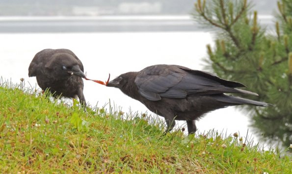
Social play involves interactions between birds – there’s play chasing, where one bird follows another, whether on the ground or through the air. We also have play fighting, where two birds engage in actions adopted from antagonistic behaviors. There’s also play invitation, where two birds engage in actions that occur primarily before play. Finally, we have social object play, where two birds engage with one or more objects in their environment (Bond & Diamond, 2003).
While we briefly touched on it, object play itself can be considered another major category of play – involving exploration, curiosity, object manipulation, where birds learn about the structure of objects, if they’re edible or not, and how they might work (Emery & Clayton, 2015).
The problem with researching play is that birds don’t necessarily need a reason to play – even when considering the anthropomorphic projection we cast onto them from our human perspectives, it can be as simple as having fun!
“Fun”, at least, in the context of the brain, involves taking part in something rewarding that provides a sense of pleasure. You may have heard of the neurotransmitter dopamine… it plays an important role in the reward-seeking system, controlling the search for reward-inducing stimuli for almost all creatures (Emery & Clayton, 2015).
When people come across phenomena like this in the wild, the answer that tends to come to mind often lies on the justifying end of the spectrum; in our attempt to be objective, we may implicitly assign a “value” to that act of play. Perhaps birds play fight to practice hunting, perhaps birds engage in social play to form strong, interconnected networks, and perhaps that crow slid down that roof to learn how to use that lid!
Studies done on the potential concrete benefits of play – such as relative brain size, for instance – ultimately find that there is little to no predictive value between the complexity of play with those benefits. As such, it is hard to say whether or not the act of play is utilitarian in nature (Bond & Diamond, 2003).
Does that mean that play is inherently fun? Once again, it’s hard to say! Humans, after all, may be the only species that have the luxury of leisure time. The time we spend solely pursuing pleasure: games, hobbies, activities we may find fun, are all a product of modern developments. We no longer have to hunt or grow our own food, we no longer have to worry about predators, we no longer have to worry about forming civilized societies – but birds, and by extension, all other animals, do (Emery & Clayton, 2015)!
Perhaps that is the underlying reason why we believe play has to have a purpose. Wild animals have a limited time to find food, find water, avoid predators, find a mate, raise offspring, all in the name of the cycle of life. Why, then, would a bird waste its time playing for the sake of playing? Why, then, do we have examples of crows sledding through the snow?
All of these questions are interesting to ponder, and I personally don’t have all the answers. The realm of neuroscience is far out of my understanding, but I hope I’ve done a good job setting the stage for this interesting question.
I’ll leave things here with a plan of action to you, the reader – whenever you see a cute animal video in the future, think to yourself: why could this be happening? Is it play, or is it intentional? Most importantly, though… why is it so gosh darn adorable?!
Reference List
Bond, A., & Diamond, J. (2003). A Comparative Analysis of Social Play in Birds. Behaviour, 140(8-9), 1091–1115. https://doi.org/10.1163/156853903322589650
Emery, N., & Clayton, N. (2015). Do birds have the capacity for fun? Current Biology, 25(1), R16–R20. https://doi.org/10.1016/j.cub.2014.09.020
I’m willing to bet you’ve never heard of Organoids. No worries if so, they’re a pretty niche part of biomedical sciences. This is such a shame however as they’re having a great impact on research and our world. Organoids are often defined as tissue samples embedded into 3D extracellular matrix (ECM) gels or clonal derivatives of cells grown without mesenchyme and derived from stem cells. Don’t worry if your eyes have already glazed over! Mine did too the first time I read that.
In simpler terms, they are the growing of cells, taken from a patient, in a nutrient rich gel. While the field might seem niche at first, it has a long history and the potential to impact millions.
Before Organoids, most human physiological research relied on in vitro 2D cell culture and animal models (O’Connell & Winter, 2019). These often fail at clinical trials due to oversimplification of or incompatibility with human systems. At the time, cell cultures were limited to growing single layers of cells in a petri dish with none of the 3D complexities of real biological systems. This all changed with Ross Harrison’s “Hanging drop Cell Cultures” in 1906 (Simian and Bissell, 2016). By creating a 2D cell culture and turning the petri dish upside down, gravity would cause the cells to clump together and form a cohesive 3D spheroid of cells that could intermingle and interact like normal. Think about that! 3D structures could form by simply turning them upside down with no other interference. This was the start of research into organoids which offered a new way to look directly at human biology without risking patient or animal lives.
One key breakthrough was the discovery of 3D extracellular matrices first used in 1963 (Simian and Bissell, 2016). The gel provided a medium for natural scaffolding of cells rather than relying on gravity. This allowed for more complex 3D shapes and allowed for more types of organoids that more closely resembled complex real life systems. The gel also aided in providing the cells with nutrients required to keep them alive. Hence the “nutrient rich gel” from the definition above.
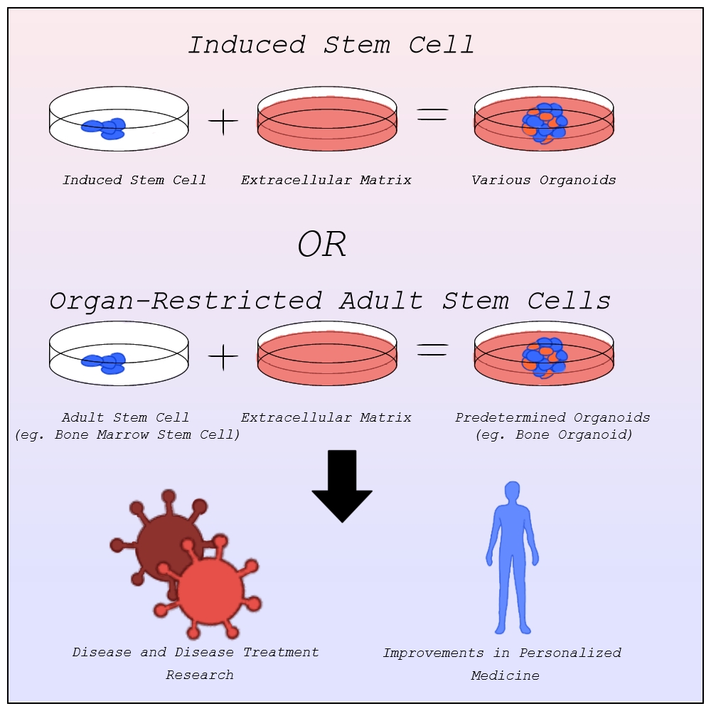
Exploration into this new field was rather slow however. The human body is composed of roughly 200 distinct types of cells, each of which specialize in a different function such as blood or nerve cells. Despite this, organoids mainly rely on only two types of cells, pluripotent stem cells and organ-restricted adult stem cells (Bredenoord et al., 2017). Stem cells are unspecified types of cells, yet to differentiate, with the ability to become any type of specified cell. They often serve to repair the human body as they can replace any damaged cells no matter the type. This ability makes them highly valuable to organoids trying to model systems that rely on the cooperation of multiple cell types.
Before the discovery of induced stem cells in 2006 by Dr. Shinya Yamanaka at Kyoto University, organoids were made from pluripotent stem cells as organ-restricted adult stem cells can be hard to access and are restricted to becoming cell types of their tissue of origin most commonly found in embryos (Agarwal et al., 2024). The line of when a zygote becomes a person, however, is blurry, raising many ethical concerns about the desecration of a person’s body. Induced stem cells allow for the change of specialized cells back into stem cells. Organoids can now be created from any tissue sample, entirely limiting the need for embryonic stem cells outside of studying early organ development (Frans Schutgens & Clevers, 2019).
With better ways to develop organoids, scientists quickly realized their potential. Organoids offer unparalleled advancement in disease research and treatment, personalized medicine, and so much more that we simply don’t have time for here.
Image by Alexandra Marshall
Disease Research and Treatment
One pivotal area of applications is understanding the development of diseases and disease treatment in humans. The lack of 3D structure in in vitro models lead to limited complexity and physiological relevance when studying diseases. Faulty predictions based on these studies can lead to treatments failing, with approximately 90% of clinical study drug candidates failing clinical trials and drug approval (Sun et al., 2022).
Organoid models have none of these limitations. In 2013, the first human condition modeled using organoids, cystic fibrosis, was published. The model had “excellent predictive value” and became the first successful personalized treatment method for cystic fibrosis patients (Lancaster and Huch, 2019). The modeling of other diseases using organoids quickly followed from genetic diseases like Polycystic kidney disease and Alagille syndrome, to infectious diseases like Covid-19 and the Zika virus, to neurological diseases like Alzheimer’s and Parkinson’s, to multitudes of cancers, and more (Xu et al., 2018). The ability for organoids to be made of any human system means they can model any disease regardless of which systems they afflict.
Organoids can be also grown co-culturally with bacteria allowing for bacterial infections and the diseases that arise from them to also be modeled and researched with organoids (Schutgens and Clevers, 2020). This has allowed for better understanding of bacteria like Helicobacter Pylori and Salmonella, aiding in detection and cures. As mentioned above, treatments can then be tested on these organoid systems without risking human lives or animal cruelty.
Respiratory Syncytial Virus (RSV), a disease that is being modeled in airway organoids, causes around 66,000–199,000 deaths among children annually (Schutgens & Clevers, 2020). This is just one of the many diseases for which scientists are now better equipped to find viable treatments. Organoids have already saved millions of lives and their potential for future disease treatment has the potential to millions more.
Personalized Medicine
While these treatments work for many people, genetic and biological differences can impact treatment effectiveness and tolerance with some even leading to negative side effects (Breen, 2021). In such situations, personalized medicine that is specialized to fit these differences might be necessary. Personalized medicine, sometimes called precision medicine, challenges the idea that treatments that pass general trials will work for everyone, instead working to construct a treatment plan with a specific patient in mind best suited to their physiology.
The idea of personalized medicine has been around for thousands of years, using one’s family’s history of diseases to diagnose the likelihood of that person having said disease. In the late 1900s the idea of evidence based medicine, the implementation of explicit reasoning based on a specific patient’s data, started being common practice when prescribing a patient (Visvikis-Siest et al., 2020). This opened the door for personalized medicine in modern science (Visvikis-Siest et al., 2020). This data was largely based on patients’ reactions to other treatments and family history however limited its effectiveness. The development of molecular biology and human genome mapping allows for a direct look at a patient’s physiology, aiding medical professionals in determining the biological reasons behind potential reactions to treatments before administration limiting adverse reactions.
Organoids have greatly furthered this field. As mentioned above, organoids are made from a patient’s induced stem cells. This means that the organoid culture into a structure based on the patient’s DNA, containing all the same biomarkers and physiology that the patient would have at that level. Where other treatments could have varying effects from the tested population, organoids allow medical professionals to see the exact effects a treatment plan would have on the patient. The use of stem cells also allows organoids to grow much quicker than other models. This allows for quick and accurate testing of the effects of medication on a patient. One study published in Science involving gastrointestinal cancer therapies found that organoids would correctly detect when drugs would fail 100% of the time, and when the drug would succeed 88% percent of the time (Vlachogiannis et al., 2018). Once again, because of the diversity of systems that organoids can model, organoids can be used to model many different personalized medicine plans allowing for the variety of treatment testing needed to find the most optimal plan.
Conclusion
Organoids have had a monumental, yet widely unknown, impact on a wide variety of disease advancement and treatment developments and personalized medicine. These structures can then be used to model human tissues and organs with much higher accuracy and complexity than previously used non-human or in vitro models.
Scientists use them to model disease development in humans and treatments designed for humans. This allows for a better understanding of the diseases and more effective treatments with fewer errors. They are also used to limit the risk of negative side effects from non-personalized medicine by furthering personalized medicine through the accuracy and speedy testing of many different treatment options.
With continued and careful development of organoids, disease understanding and treatments, personalized medicine and so much more can continue to be improved, saving millions of lives.
————————————————————————————————-
Work Cited:
Agarwal, S., Saha, L., Sohani, A., Karri, U., Gor, R., & Ramalingam, S. (2024). The importance of niche in therapeutic strategies targeting normal and cancer stem cells. Cancer Stem Cells and Signaling Pathways, 15–33. https://doi.org/10.1016/b978-0-443-13212-4.00031-3
Bredenoord, A. L., Clevers, H., & Knoblich, J. A. (2017). Human tissues in a dish: The research and ethical implications of organoid technology. Science, 355(6322). https://doi.org/10.1126/science.aaf9414
Frans Schutgens, & Clevers, H. (2019). Human Organoids: Tools for Understanding Biology and Treating Diseases. Annual Review of Pathology Mechanisms of Disease, 15(1), 211–234. https://doi.org/10.1146/annurev-pathmechdis-012419-032611
Generation and Cultivation of 3D Cell Models | ibidi. (2024). Ibidi. https://ibidi.com/content/1037-generation-and-cultivation-of-3d-cell-models
Lancaster, M. A., & Huch, M. (2019). Disease modelling in human organoids. Disease Models & Mechanisms, 12(7). https://doi.org/10.1242/dmm.039347
O’Connell, L., & Winter, D. C. (2019). Organoids: Past Learning and Future Directions. Stem Cells and Development, 29(5), 281–289.https://doi.org/10.1089/scd.2019.0227
Schutgens, F., & Clevers, H. (2020). Human Organoids: Tools for Understanding Biology and Treating Diseases. Annual Review of Pathology: Mechanisms of Disease, 15(1), 211–234. https://doi.org/10.1146/annurev-pathmechdis-012419-032611
Simian, M., & Bissell, M. J. (2016). Organoids: A historical perspective of thinking in three dimensions. The Journal of Cell Biology, 216(1), 31–40. https://doi.org/10.1083/jcb.201610056
Sun, D., Gao, W., Hu, H., & Zhou, S. (2022). Why 90% of clinical drug development fails and how to improve it? Acta Pharmaceutica Sinica B, 12(7), 3049–3062. https://doi.org/10.1016/j.apsb.2022.02.002
Visvikis-Siest, S., Theodoridou, D., Kontoe, M.-S., Kumar, S., & Marschler, M. (2020). Milestones in Personalized Medicine: From the Ancient Time to Nowadays—the Provocation of COVID-19. Frontiers in Genetics, 11. https://doi.org/10.3389/fgene.2020.569175
Vlachogiannis, G., Hedayat, S., Vatsiou, A., Jamin, Y., Fernández-Mateos, J., Khan, K., Lampis, A., Eason, K., Huntingford, I., Burke, R., Rata, M., Koh, D.-M., Tunariu, N., Collins, D., Hulkki-Wilson, S., Ragulan, C., Spiteri, I., Moorcraft, S. Y., Chau, I., & Rao, S. (2018). Patient-derived organoids model treatment response of metastatic gastrointestinal cancers. Science, 359(6378), 920–926. https://doi.org/10.1126/science.aao2774
Xu, H., Jiao, Y., Qin, S., Zhao, W., Chu, Q., & Wu, K. (2018). Organoid technology in disease modelling, drug development, personalized treatment and regeneration medicine. Experimental Hematology and Oncology, 7(1). https://doi.org/10.1186/s40164-018-0122-9
Details from my summer experience conducting fieldwork in the Colorado Rockies
Photo caption: View of the Rocky Mountain Biological Laboratory, with Gothic Mountain towering above.
4:30 AM: Good morning! To arrive at my research sites in time to start my bird species surveys by 6 AM, I often have to wake up long before the sun rises. I get dressed, refill my water bottles, collect the snacks and PB&J sandwiches I prepared the night before, and grab my pack. Inside my pack I have my binoculars, data collection sheets, a handheld GPS unit, and of course a first aid kit and extra warm layers. Field research requires that you always be prepared for the unexpected!
This summer I am investigating how birds in the southern Rocky Mountains are responding to climate change. To do this, I am conducting a resurvey of bird species along an elevational gradient. I have six research sites ranging from 8,100-11,200ft in elevation, and I will visit each site three times throughout the season to conduct a point count survey of bird species. A point count survey involves standing in one spot for a specific amount of time and recording all bird species I observe by sight or sound. Another student conducted these surveys in 2011, so I am repeating their methods to be able to compare the data from 2011 and 2023 and examine how bird communities have changed over the 12-year period.
6:00 AM: After biking 3 miles along a gravel road to the base of Snodgrass Mountain and hiking another 3 miles up the slope (including a break halfway to enjoy my PB&J for breakfast), I am ready to begin the point counts. I use the GPS unit to locate the coordinates of each survey point, and record all of the species that I hear and see during a five-minute period. These surveys require me to be very familiar with the songs and calls of bird species in the region, as the majority of my detections are auditory. I audio record all of my point counts so that I can go back and verify observations later if I am not certain about my real-time identification in the field.
8:00 AM: After I finish the point counts, I sit down to eat a snack and enjoy the beautiful views around me. I begin my bike ride down the mountain, whizzing through fields full of blooming wildflowers.
Photo caption: Views of the sunrise and wildflowers from my trek up Snodgrass Mountain.
9:30 AM: I make it back to my home for the summer: the Rocky Mountain Biological Laboratory (RMBL), a thriving community of almost 200 field biologists, each studying one component within the greater ecosystem of the southern Rocky Mountains, united with the goal of recording long-term ecosystem responses to climate change. I head to the dining hall to grab more snacks and refill my water—field work can be exhausting and it’s especially important to stay hydrated.
10:00 AM: I head back to my cabin to enter my point count data from the morning. This involves transcribing my handwritten field notes on paper datasheets into a spreadsheet on my laptop. This is important for multiple reasons: it serves as a backup in case my paper datasheets are lost or damaged, and formatting an electronic file is the first step to performing statistical analyses to uncover trends and relationships in one’s data.
11:00 AM: Now it’s naptime! With a wake-up call before sunrise, I almost always need a nap to recharge after morning fieldwork.
12:00 PM: I nap until lunchtime, and then I head down to the dining hall. RMBL has a great kitchen staff that makes sure the researchers are well fueled for their fieldwork, and the dining hall is a perfect place to meet other scientists and learn about all of the cool research taking place.
1:00 PM: After lunch, I take some time to look over my research report draft and make some edits to the methods section because a couple of aspects have changed since I began my surveys out in the field. Keeping good records on the methods used for surveys and experiments is very important because it ensures future researchers will be able to reproduce the experiments.
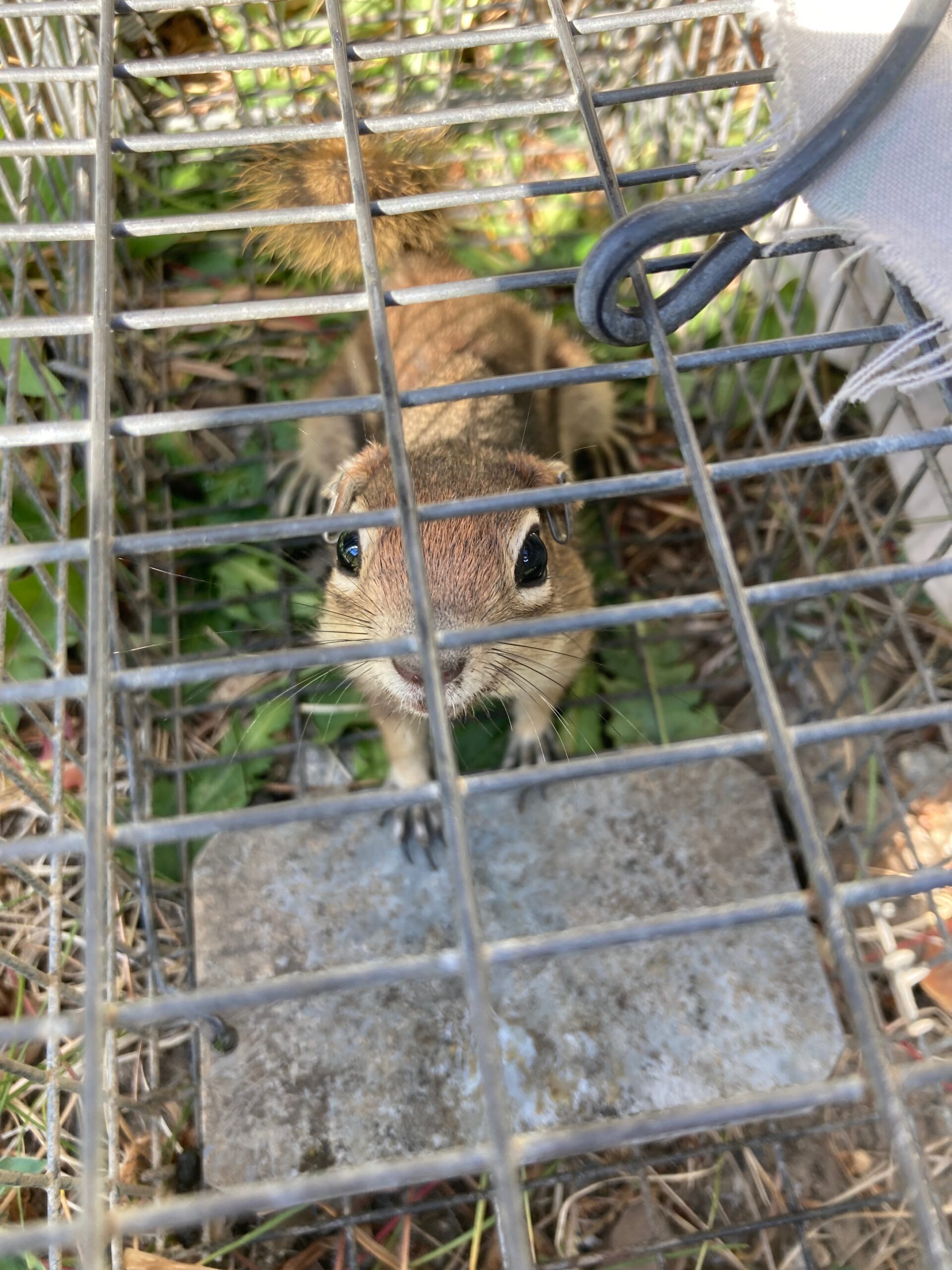
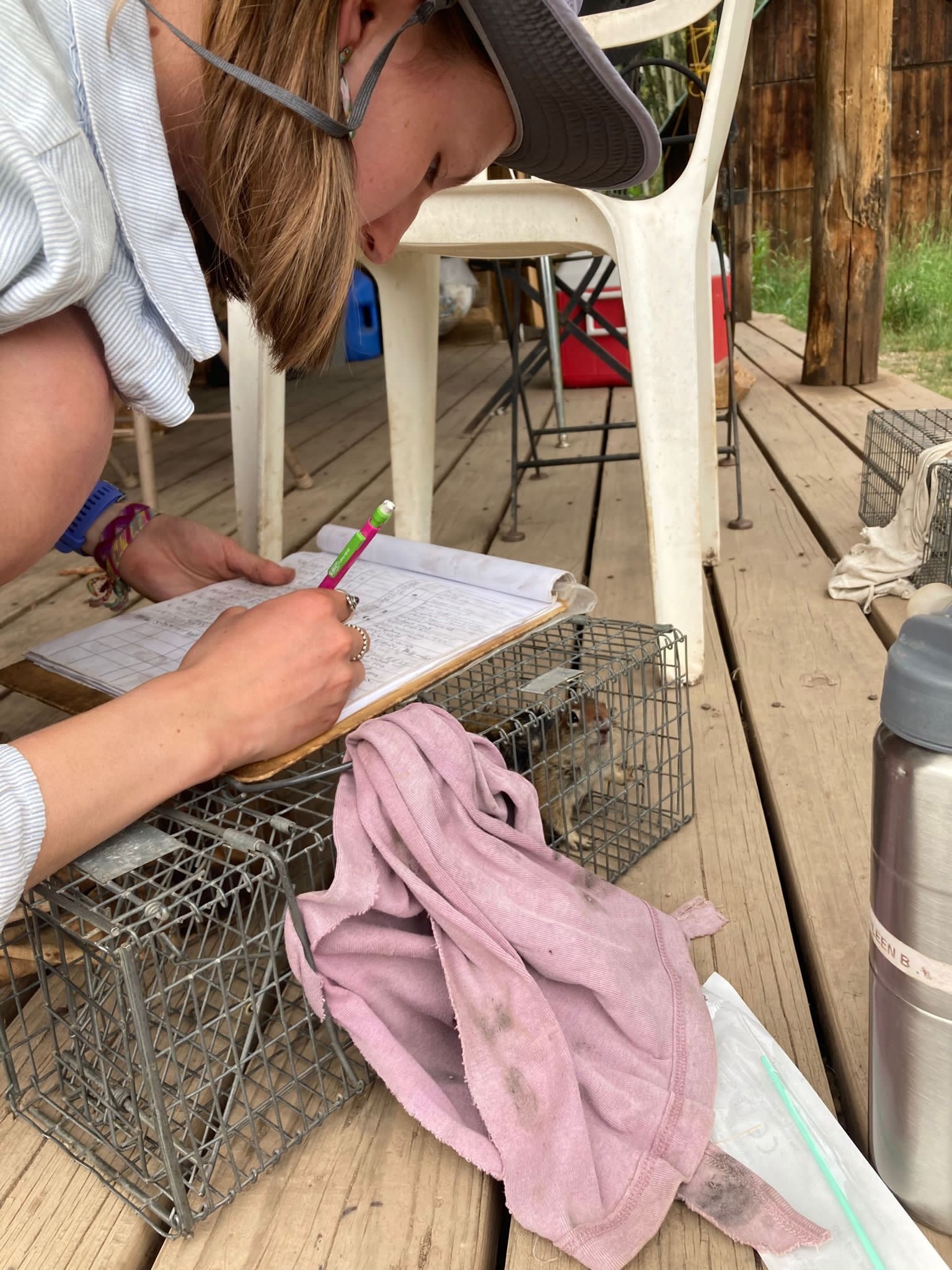
2:00 PM: Most afternoons, I work as part of the golden-mantled ground squirrel (GMGS) team on a long-term behavioral and spatial ecology project. This involves making rounds around the RMBL townsite looking for squirrels and recording their behavior in 10-minute periods. GMGS resemble chipmunks, but they are larger and can run faster. Another big component of the squirrel project is live trapping, where we bait small wire traps with peanut butter and place them around known squirrel territories. When we capture a squirrel, we put them into a special canvas bag for safe handling and we record morphological measurements such as weight and take hair samples for genetic analysis. The most exciting time is mid-summer when the squirrel pups emerge from their burrows. We try to trap pups every couple of weeks to monitor their growth.
From left to right: A GMGS inside a trap, data collection during GMGS trapping.
5:00 PM: After my shift with the squirrel team, I head back to my cabin to rest and relax until dinner. I also must pack up my camping gear, because the site I am surveying tomorrow morning is several miles up the valley from the field station, and it requires an overnight backpacking trip to get there. I pack a sleeping bag, sleeping pad, small 2-person tent, extra layers, water filter, and my survey equipment in a backpacking pack. At the dining hall during dinner, I will grab extra snacks and make a couple of PB&Js to carry with me.
7:00PM: After dinner, myself and two of my fellow student researchers borrow a vehicle to drive to the beginning of the hiking trail that will take us to the research site. We park next to a beautiful alpine lake and begin the 3-mile hike. We will gain 1500 ft in elevation over the course of the hike, so the majority of the trail is unrelentlessly steep. The gorgeous views of the sunset over the mountains around us serve as a good distraction from the strenuous hike. Two miles into the hike, we reach a river crossing. We take off our hiking boots and socks and roll up our pants to wade through the ice-cold water. Once across the river, we know there’s only a mile left to climb, and the sunlight is fading fast so we push onwards to try and make it to the site before dark.
Once we get to the site, which consists of a series of ponds located along a rocky mountain slope, we quickly set up our tents and get ready for bed. The stars are incredible here—we can even see the outline of the milky way.
Left: View of Emerald Lake from trail above. Right: Sunset over the valley after the river crossing.
Photo caption: View down into the river valley at twilight.
9:30PM: Time for bed! After the sun sets, it gets very cold at this high elevation, and the most comfortable place to be is inside my warm sleeping bag. I set my alarm for 5:30AM and make sure all of my survey equipment is ready for the morning.
This summer I learned a lot about how to effectively design and conduct a field ecology research study—especially how to be flexible and creative when problems arise. Field research is challenging but it is so refreshing to work outside in such beautiful places, and it’s especially rewarding to explore the trends in one’s data after a season of hard work. That’s how I’ll be spending the winter months, as do many field ecologists outside of field season—crunching the numbers in order to reveal the mechanisms of change in the ecosystems around us.
The effort to bring greenspace and restore natural ecology to the Georgia Tech campus can be seen in the EcoCommons, a park built near the Keneda building and the Center Street Apartments. One of the goals of the EcoCommons is to restore Georgia’s natural plant life to Atlanta. Today, we’re going to look at some of the native plants which have been brought back to the EcoCommons.
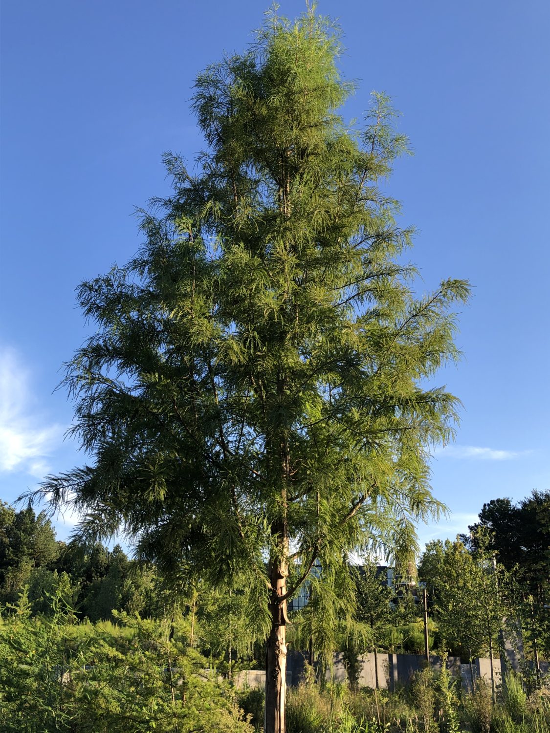
Bald Cypress Trees
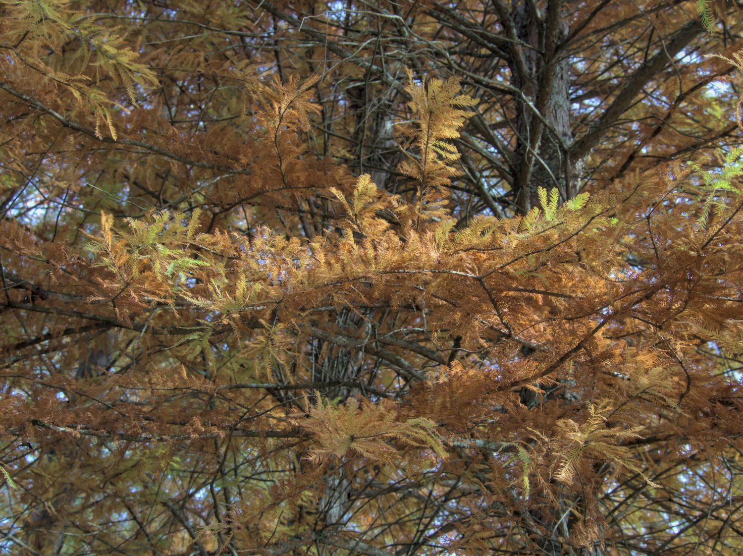
Bald cypresses are trees native to much of the southeastern United States. They’re hardy and versatile, and can grow in all kinds of soil environments. This, along with their lovely red and orange needles during the fall, makes them popular plants for decorative purposes. Other common names for the bald cypress include swamp cypress, white cypress, tidewater red cypress, gulf cypress and red cypress.
In the wild, bald cypresses are typically found in wet, swampy areas. They can be identified by their large, spread out needles.
FUN FACTS
The oldest bald cypress tree (that we know of) has been alive since 605 BCE. That’s a 2627 year old tree!
The site of the tree is actually a secret – park rangers don’t want visitors vandalizing it.
The bald cypress is the state tree of Louisiana.
Bald cypress wood has historically been marketed for its resistance to water, which makes it useful in wet areas.
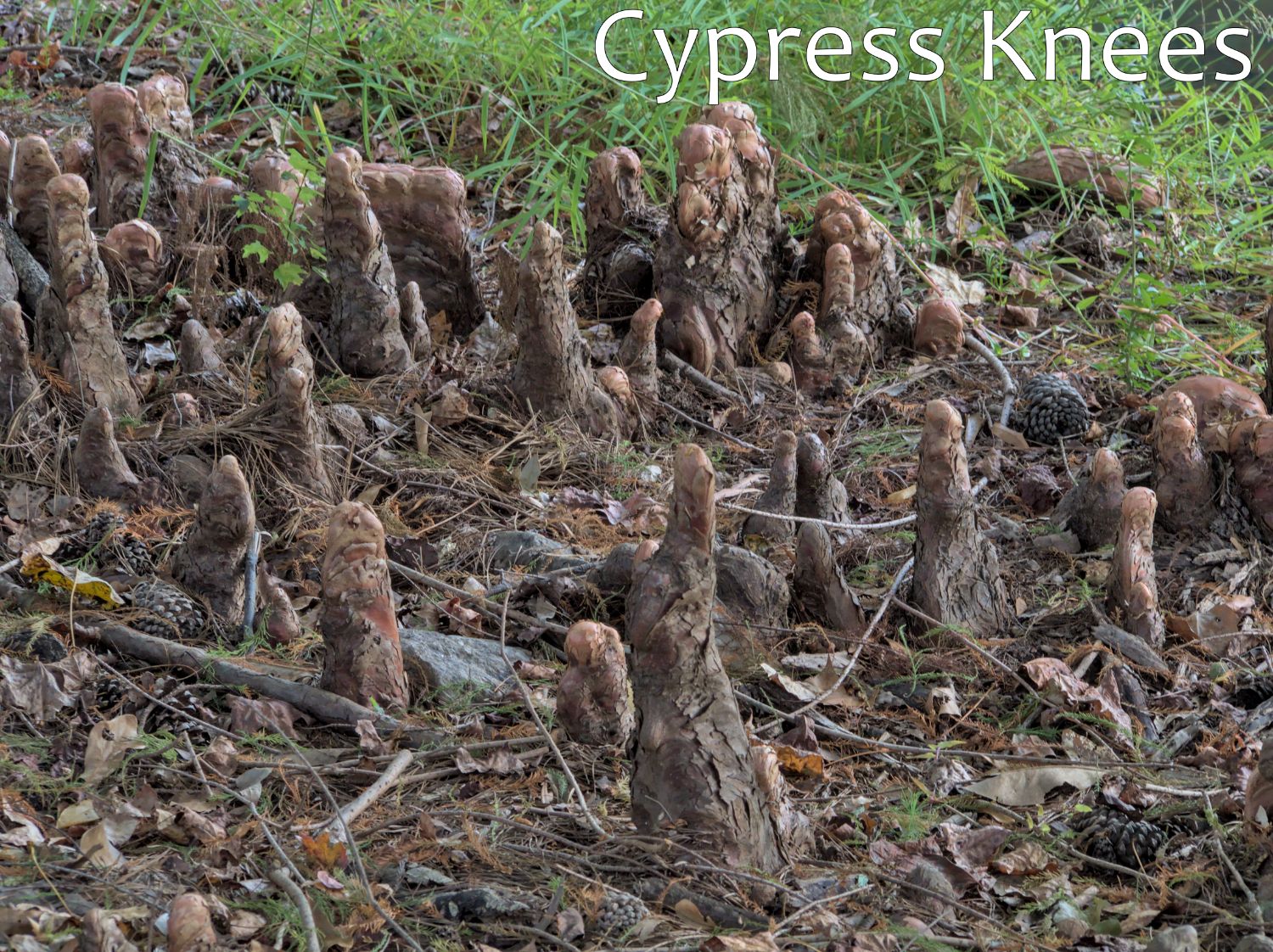
Around a bald cypress tree, it’s common to see what are called cypress knees – wooden outgrowths from the ground. They actually extend upward from the main trunk’s root system. They look like small stumps coming out of the ground, although they’re still connected to a “main” tree.
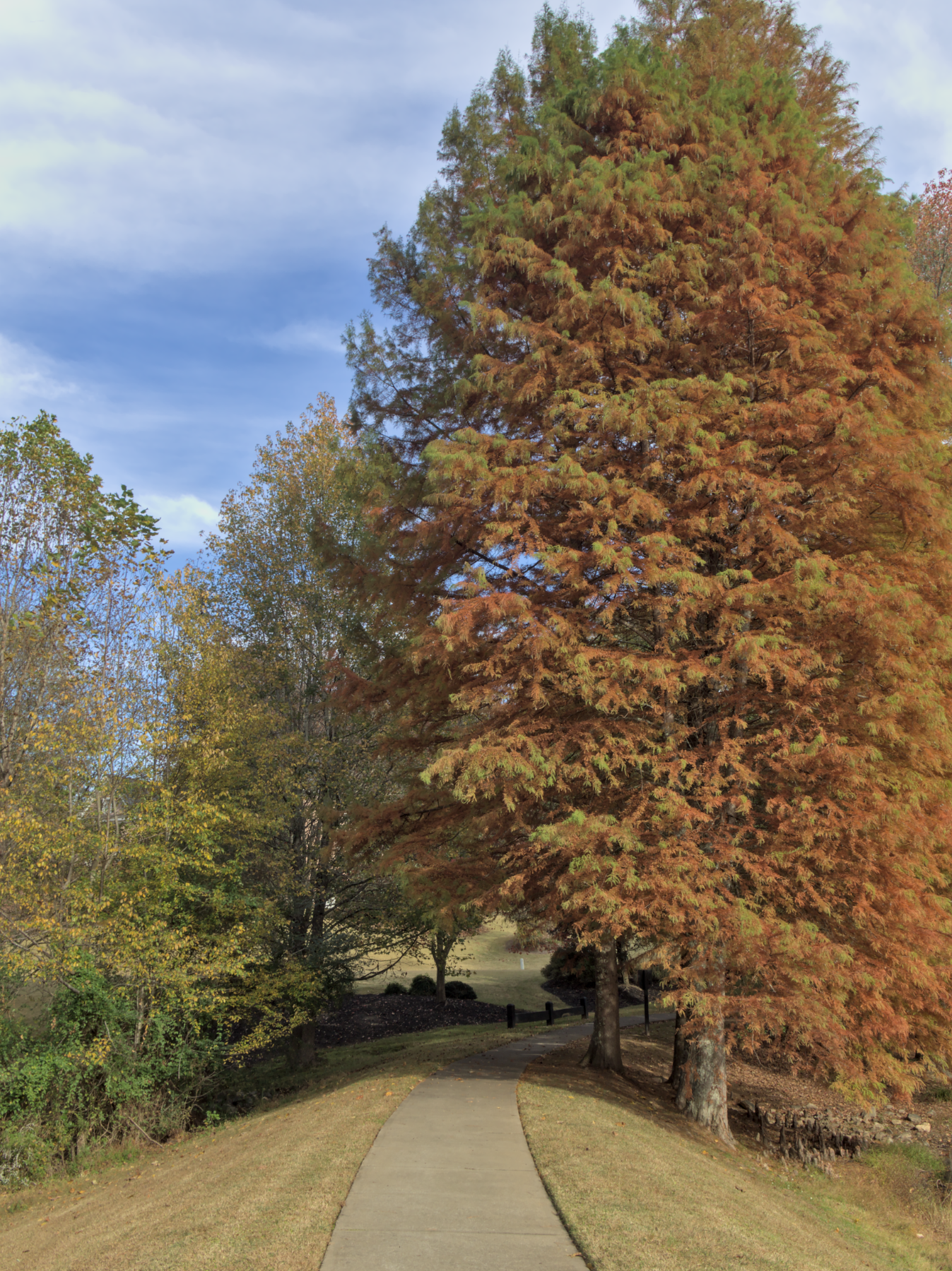
Bald cypress trees are famous for their fall colors – here’s a larger bald cypress off of the Georgia Tech campus in November. These native plants are a great choice for people looking for decorative trees that are ecologically beneficial to the local environment.
Cardinal Lobelias
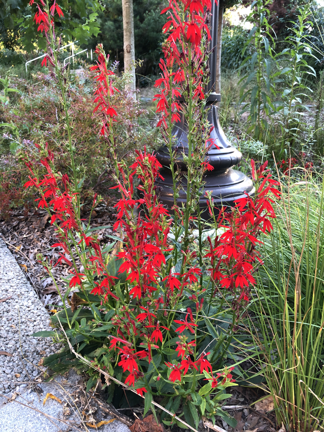
The cardinal flower, lobelia cardinalis, is a red flower, belonging to the bellflower family, that is native to most of the United States (except for far Northwestern states). It primarily lives in moist areas, such as stream banks, ditches, and lake shores.
FUN FACTS
Excellent for hummingbird and butterfly gardens. This plant attracts many native pollinators, so it’s a great addition to your yard or garden if you’d like to see more hummingbirds and butterflies.
You can find varieties of the cardinal flower with pink and white flowers, although these are mostly sold in nurseries for decorative plants.
Every part of the cardinal flower is poisonous when ingested – don’t eat it!
New England Aster
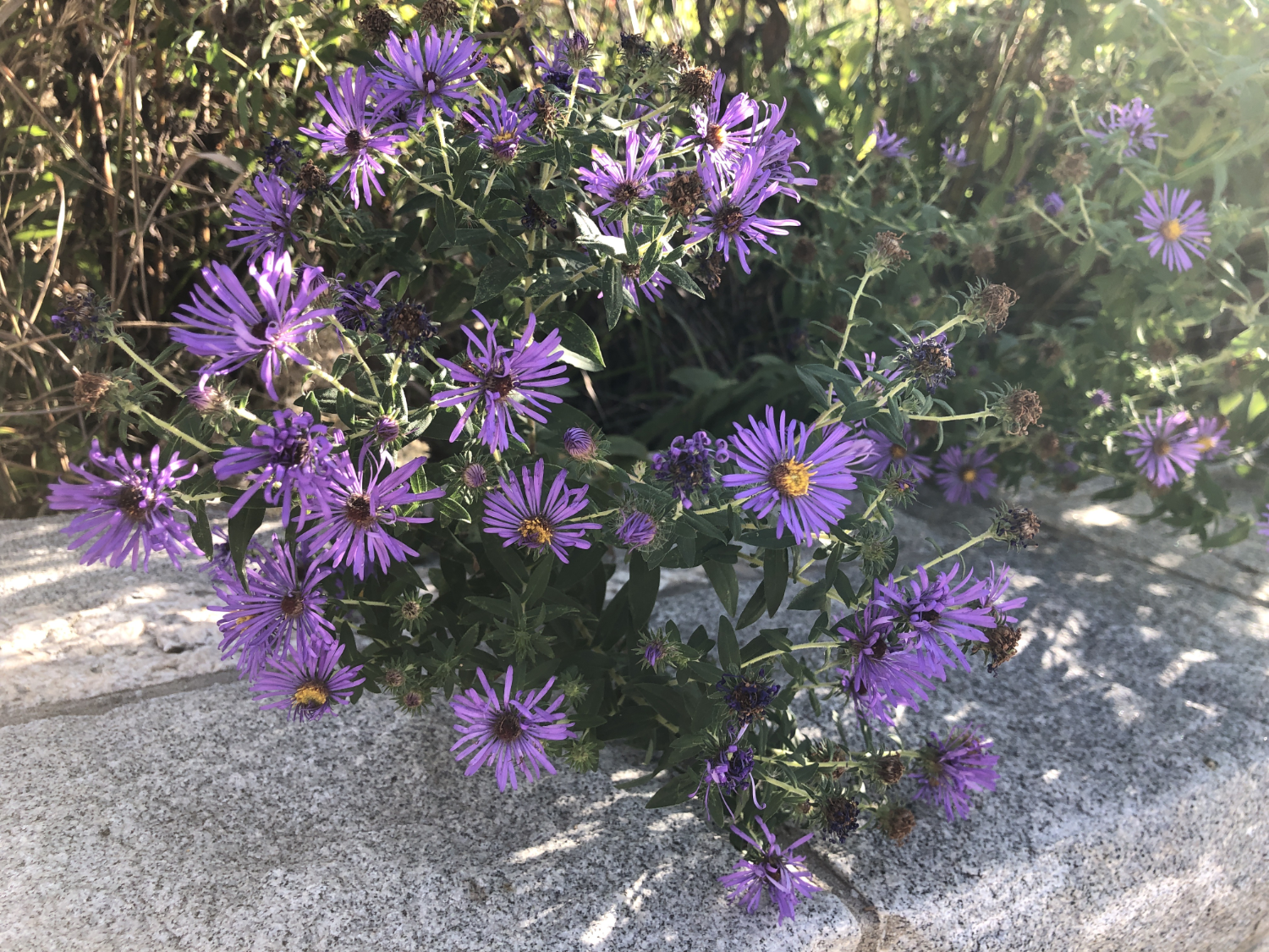
The New England aster, Symphyotrichum novae-angliae, is a perennial flower that is a native plant to much of the northeastern United States. It typically blooms from late summer through fall, with asters blooming in late August to early November depending on the region. New England asters are typically found in moist environments, particularly in wetlands. However, it has been found in dry soils as well.
FUN FACTS
New England asters are a favorite flower of the monarch butterfly! These flowers are known to attract the monarch butterflies during their migration south for the winter.
The word “aster” comes from the Greek word ἀστήρ, meaning “star”. Aster shares a language history with astronaut and astronomy.
Here in Georgia, New England asters are considered “Critically Imperiled” by NatureServe – this means that their natural range has been reduced significantly by human activity.
Domestically, you can find New England asters in all sorts of pinks and purples at nurseries for your garden or yard.
Blue Mistflower
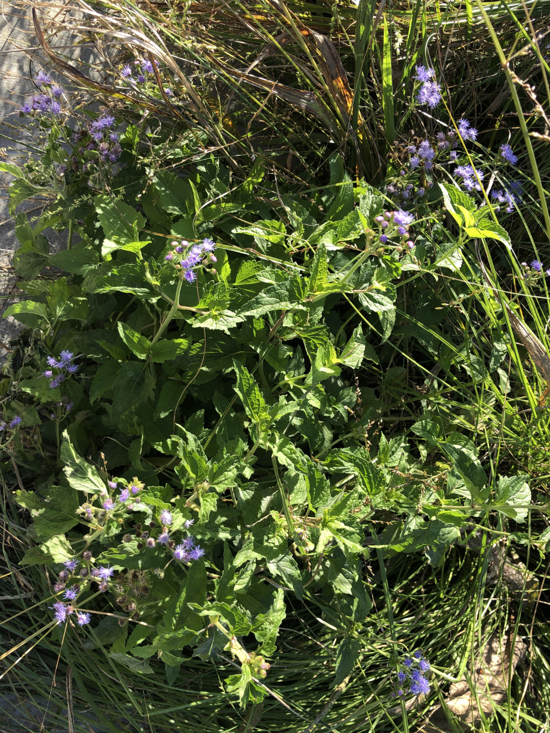
The blue mistflower, conoclinium coelestinum, is a perennial flowering plant in the Asteraceae family. Blue mistflowers are widespread throughout North America, and can be found from Ontario, Canada to Tepoztlan, Mexico. It’s commonly grown in gardens, and flowers from July through November each year.
FUN FACTS
Blue mistflowers are hardy plants that can spread quickly – be warned if you want them in your garden! They’re very capable of choking out their neighbors because their root systems spread out.
These flowers are known for their ability to attract pollinators, which makes them great for supporting local ecology.
You may have heard that modern birds evolved from dinosaurs before. More specifically, modern birds descend from theropods, which is a clade of dinosaurs. A clade is a group of species that share some key traits as well as common ancestry. Theropods all had hollow bones and three toed limbs, traits that remain in modern birds. The most famous of theropods was the T. Rex, but you also may have heard of the Spinosaurus, Archaeopteryx, and Allosaurus.
How did we come to this conclusion? It turns out hollow bones and three toed limbs are not the only similarities between theropods and modern birds. Various types of theropods have also been found to have feathers in fossilized remains. Older theropod species, that are “farther” back from dinosaurs on the tree of life, only had one kind of feather, which paleontologists think was used for insulation. Younger theropods, more closely related to birds, such as the oviraptors, had multiple different kinds of feathers. While some of these feathers are also for insulation, others are very similar to the flight feathers on modern birds. Modern birds have three “fingers” in the bone structure that supports their wings. Groups of dinosaurs that are theorized to be closely related to modern birds tend to have less and less fingers. The Orinsthican dinosaurs, very distant ancestors to birds, had five fingers. The Allosaurids, closer ancestors, had four fingers. Eventually, dinosaurs like Archaeopteryx had three fingers, which would eventually lead to the development of modern birds’ wings. Over time, the tails of dinosaurs began to get shorter and shorter. Eventually, the tails got so short that they’re barely there, until the only modern evidence of them are the tail feathers on the backs of birds.
References
Agnolin, F. L., Motta, M. J., Brissón Egli, F., Lo Coco, G., & Novas, F. E. (2019). Paravian phylogeny and the dinosaur-bird transition: An overview. Frontiers in Earth Science, 6(February), 1–28. https://doi.org/10.3389/feart.2018.00252
The authors of “Enriched environment exposure accelerates rodent driving skills”, published in Behavioral Brain Research, conducted an experiment intended to study the impact of the environment on the efficacy of stress-releasing mental health treatments in lab rats. This study aimed to see how an enriched environment, both socially and intellectually, promotes stress relief while doing fun activities. The process for teaching the rats to drive the rodent operated vehicles (known as ROVs) also has implications for the importance of enrichment in how service dogs are trained.
Study Summary
There were two groups of rats that participated in this study. The control group of rats lived in a standard environment for laboratory rats, in standard laboratory cages, each with one other rat. The experimental group of rats lived in housing with an enriched environment. These rats got multiple rat friends, rat toys to keep them occupied, and multi-level housing.
The study began with a “training period”, where rats in both groups were taught by operant conditioning how to drive the ROVs. While the authors of the study expected the rats living in standard housing to perform worse than the other rats, they were shocked at how much worse they performed. The authors noted that the control group rats failed to perform even “basic” tasks in driving the ROVs.
After the training period (in which food rewards were used to encourage the rats to drive), both groups of rats continued to drive the ROVs even in the absence of rewards. However, rats living in enriched housing drove the ROVs without treats for a longer period of time than the rats living in standard housing. Throughout the study, the scientists measured the levels of hormones that were indicative of the rats’ ability to handle stress. Both groups showed an improved ability to handle stress as a result of ROV driving, but the enriched housing rats showed a significantly larger benefit than the standard housed rats.
Conclusion
This investigation shows that the environment of an animal has a significant impact on the effect of stress relieving interventions. The authors suspect that the enriched rats were more able to learn new behaviors (driving the ROVs) as a result of experiencing a wider variety of behaviors in their enriched home environments. The results of this study have interesting ramifications on the optimal conditions for other animals who will be undergoing rigorous training. Additionally, this study demonstrates how a good environment that has opportunities for social connection and mental challenges is beneficial in mental health treatment.
References
Crawford, L. E., Knouse, L. E., Kent, M., Vavra, D., Harding, O., LeServe, D., Fox, N., Hu, X., Li, P., Glory, C., & Lambert, K. G. (2020). Enriched environment exposure accelerates rodent driving skills. Behavioural Brain Research, 378, 112309. https://doi.org/10.1016/j.bbr.2019.112309
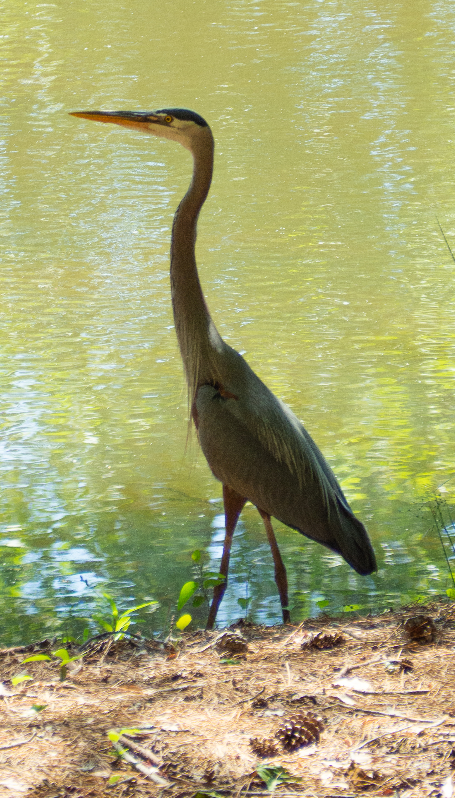
Great blue herons are part of the Ardea genus, which is a genus of birds that includes the herons, egrets, and bitterns. These birds are typically pretty large in size, with lengths usually greater than 80–100 cm. You’re most likely to find members of this genus in wetlands and other partially aquatic habitats.
Feeding Behavior
Great blue herons are carnivores, and they eat a variety of aquatic, semi-aquatic, and terrestrial prey. Their diet consists of predominantly fish, but they will eat pretty much anything that they can catch in their beaks. Fish are supplemented by amphibians, reptiles, small ground mammals, and even other birds. Great blue herons catch smaller prey in the jaws at the back of their beak, although they prefer to stab larger prey with their beaks.
There are 38 total feeding behaviors that have been recorded by members of the Ardea family. Each species in this genus uses multiple feeding methods — they’re creative hunters. Twelve of these strategies have been observed in great blue herons. The most commonly observed feeding behavior for the great blue heron is standing and waiting, where the heron simply stands still and waits for its prey to come to it. Herons also will move slowly, either through the water or through a field, in search of prey. More dramatically, the great blue heron will occasionally plunge-dive, diving down quickly and with force, to catch its prey. A particularly interesting feeding behavior of the great blue heron is to stick its bill in the water, and vibrate it. The underwater vibrations create ripples, attracting fish and other aquatic prey.
Habitat
Great blue herons prefer to live in wetlands, especially when nesting. Rather than nesting alone, great blue herons nest in colonies. Colony nesting is particularly common for birds that live in aquatic environments. Colonies can have over 500 nests (so easily over a thousand birds) all relatively close together. For large scale habitats and especially for colony nesting, great blue herons have preferences for certain types of habitats over others. The lacustrine wetlands is a particular favorite of Illinois great blue herons. Lacustrine wetlands are any wetland or deepwater area that has these three characteristics:
Located in a valley/depression in the ground
Lacking trees, shrubs, and other plant life that covers more than 30% of the area
At least 20 acres in surface area
Biologists believe that herons may prefer this biome for their habitats due to the easy access to their preferred prey of small bodied vertebrates. Additionally, large wetlands tend to be isolated from human activity, which great blue herons tend to avoid if possible.
References
Combs, B. P. (2021). Great Blue Heron Forages for Fish by Bill-Vibrating. Western North American Naturalist, 81(2), 280–284. https://doi.org/10.3398/064.081.0213
Gibbs, J. P., & Kinkel, L. K. (1997). Determinants of the Size and Location of Great Blue Heron Colonies. Colonial Waterbirds, 20(1), 1–7. https://doi.org/10.2307/1521757
Knight, E. C., Vennesland, R. G., & Winchester, N. N. (2016). Importance of Proximity to Foraging Areas for the Pacific Great Blue Heron (Ardea herodias fannini) Nesting in a Developed Landscape. Waterbirds, 39(2), 165–174. https://doi.org/10.1675/063.039.0207
The red-shouldered hawk (Buteo lineatus) is a raptor that is found in forests throughout North America. Year round, the red-shouldered hawk is found throughout much of the Eastern and Midwestern United states. During their breeding season, they can be found in both the northeastern and western United States as well as part of Atlantic Canada. They also have a non-breeding range in Mexico, where they are found for some parts of the year. Red-shouldered hawks can be identified by their medium size and reddish-brown feathers on their chests and shoulders. Hawks from the western part of their range tend to be a deeper red than those found in the eastern part. As for their wings, the feathers are banded black and brown from the underside, with black and red being predominant on the outside. Their tails are also banded, which is another identifying trait of the species.
Sexual Dimorphism in Red-shouldered Hawks
Red-shouldered hawks do show some sexual dimorphism in their appearance. Sexual dimorphism occurs when males and females of the same species show different traits on average, such as average size and weight. Mass is one of the traits that is sexually dimorphic for red-shouldered hawks. Females are larger than males, with the average male being only 85% of the mass of the average female. Researchers recently have tried to establish other ways of determining the sex of red-shouldered hawks morphologically. Determining sex by morphology is used by biologists to guess at the sex of an animal without needing to check for sure with DNA testing. Sex determination based on morphology is more convenient than determining sex based on molecular testing, and can be done by biologists in the field without access to DNA testing equipment. Other features of red-shouldered hawks that are sexually dimorphic are toepad length, length of secondary feathers, bill length, mass, and wing chord length. However, mass remains the most reliable way to morphologically determine the sex of a red-shouldered hawk.
Diet and Habitat of the Red-shouldered Hawk
The diet of the red-shouldered hawk is mostly made up of small mammals, lizards, snakes, and amphibians, and these hawks typically live in riparian forests. Riparian forests are forests that are on or near the bank of a river or stream. There is evidence that the composition of the red-shouldered hawk’s diet varies depending on where in their range they are living. One study split red shouldered-hawks into two groups. The first group was the northern hawks, which live above 38o N latitude. This is the latitude line passing just south of Charlottesville, VA, and through Lexington, KY. Interestingly, northern hawks preferred more mammalian prey and less amphibian prey. The opposite was true for southern hawks. Researchers noticed that diets were more mammalian in drier conditions, whereas hawks that lived in wetter conditions relied more on invertebrate and amphibian prey. It’s possible that hawks that live in drier areas will be less affected by wetland degradation than hawks in wetter areas.
Nesting Preferences of the Red-shouldered Hawk
Where red-shouldered hawks choose to nest is another area of interest for researchers. Typically, red-shouldered hawks prefer to nest in large tracts of mature forest, although they can also be found in more fragmented forested areas, such as those found in suburban areas. One study found that nests were more likely to be located near a lake or treeless wetland than not. Nests were also more likely to be near a trail or stream than not for both cases. Researchers believe that open trails might make good hunting grounds for the hawks, without trees and underbrush to hide prey. We can guess similarly about hawks’ proximity to lakes and treeless wetlands. The lack of trees gives hawks easier access to their prey, which they detect primarily via eyesight. Red-shouldered hawks’ positive association with wet areas is a well known feature – amphibians make an important component of the diet of the red-shouldered hawk, especially while nesting.
References
Cheryl R. Dykstra, Herman L. Mays Jr, Jeffrey L. Hays, Melinda M. Simon, & Ann R. Wegman. (2012). Sexing Adult and Nestling Red-Shouldered Hawks Using Morphometrics and Molecular Techniques1. Journal of Raptor Research, 46(4), 357–364. https://doi.org/10.3356/JRR-11-84.1
Brad N. Strobel, & Clint W. Boal. (2010). Regional Variation in Diets of Breeding Red-Shouldered Hawks. The Wilson Journal of Ornithology, 122(1), 68–74. https://doi.org/10.1676/09-071.1
Landenberger, James F, Roosa, Dean M, Stravers, Jon W, Ehresman, Bruce, & Patterson, Rich. (2013). The Raptors of Iowa. University of Iowa Press.
James E. Woodford, Carol A. Eloranta, & Anthony Rinaldi. (2008). Nest Density, Productivity, and Habitat Selection of Red-Shouldered Hawks in a Contiguous Forest. Journal of Raptor Research, 42(2), 79–86. https://doi.org/10.3356/JRR-07-44.1
Red-shouldered Hawk Overview, All About Birds, Cornell Lab of Ornithology. (2019). allaboutbirds.org. https://www.allaboutbirds.org/guide/Red-shouldered_Hawk/