Tag: medicine

  • Organoids Part Two: Despite Limitations, They’re Still Amazing

    Clearly one article wasn’t enough to explore the full potential of organoids, so here we go again. This article won’t make much sense without reading the first but a quick catch up:
    Organoids are 3D cell structures grown from Stem cell samples and grown in nutrient rich gel.

    • They can better model human systems as they are more complex than their 2D counterparts and a better representation of human systems than animal models.
    • This advantage makes organoids uniquely beneficial to the field of drug research and testing and personalized medicine.
    • Link to original article if desired: https://2026.chargedmag.org/2026/04/organoids-what-are-they-and-why-theyre-amazing/

    Limitations
    These applications are not without limitations however. While organoids can replicate many aspects of human tissue, some specific cell genes are harder to find in organoids (Andrews and Kriegstein, 2022). This impaired the organoids’ ability to properly simulate the systems they are derived from, preventing researchers from properly modeling diseases and treatments in that area.

    Additionally, while developments have been made to improve organoid lifespans, organoids still require controlled environments and regular care to survive (Simian and Bissell, 2016). Even the longest lasting organoids, neurological models, have only survived a couple of years. Many organoids will even grow patches of dead tissue in the center due to a lack of oxygen hindering their research capabilities (Andrews & Kriegstein, 2022). In organic systems, cells are constantly interchanging molecules to maintain their functions and prevent necrosis. The nutritional gel can only do so much to get the molecules to cells at the interior of organoid structure without the natural systems to supply it.

    All of this causes organoid maintenance to be costly compared to in-vitro models, which are relatively cheap and easy to handle (Schutgens & Clevers, 2020). This stifles research leading to many knowledge gaps in the field. Furthermore, these restrictions limit modeling larger systems. Potential treatments tested in smaller organoids could have unpredictable impacts on a larger system when prescribed. Concerns around this limits applications of organoids, creating Theory-Application Voids.

    There are also many population gaps in organoid research as certain demographics are underrepresented in the stem cells used to create the organoids. These stem cells are often pulled from stem cell donor registries which have significant demographic, systemic, and behavioral biases. Further development into organoids and increased diversity in biobanks can increase organoid capabilities and applications.

    Concerns
    There are also current concerns surrounding organoid research that need to be addressed. While organoid models no longer rely on stem cells taken from embryos, they are still used in organoids modeling traditional embryo frameworks. These models use STEM cells to study the process of early embryonic development and can be very useful in identifying gestation concerns or potential risks to a fetus (Pereira et al., 2020). Once again, the line of when a zygote becomes a person however, gets blurry. Multiple public figures and many private citizens are debating if the creation of an embryo, even as a limited organoid, for study is ethical.

    There are also concerns about neurological organoid models used for neurological diseases (Lavazza and Massimini, 2018). Current models can only imitate parts of the human brain. However, as models more closely resemble a human brain with increased functionality, ethical questions surrounding the creation and experimentation of something than can think and feel will only grow. The line being living, sentient, and sapient is hard to discern, especially with limited structures that might not have the ability to communicate mental facilities.

    Future implementations of organoid models also raise ethical concerns regarding their financial exploitation and patient consent. People’s cell matter must be collected and added to databases to be used in organoid research (Bredenoord et al., 2017). Active patient involvement in organoid development is essential when considering this process. Their participation in cell donation must remain voluntary and properly compensated, and this is currently not guaranteed.

    The implementation of organoids in personalized medicine could also have complications on the healthcare system (Stefanicka-Wojtas & Kurpas, 2023). While organoids could improve personalized medicine, the increased specificity of treatments could lead to decreased procedure lifespans and certainty. The underrepresentation and price of organoid backed research could also exacerbate existing disparities in healthcare and lead to an increase in general healthcare costs or dropped plans due to cost-shifting (What’s Behind Rising Health Insurance Costs? | Johns Hopkins Bloomberg School of Public Health, 2025).

    Scientists in organoid research have repeatedly advocated for remaining cautious and careful in this relatively unexplored ethical field.

    Potential Applications
    Despite all of that dour news, organoids are still having a positive impact on millions of people with the potential to aid millions more. Unfortunately we only have time for two of these here: Regenerative Medicine and Transplantation.

    Regenerative Medicine is the field of science dedicated to the regeneration and restoration of human tissue based in Stem cells. Many different animals have regenerative properties from the regrowth and axolotl limbs to sea cucumbers regrowing internal organs to flatworms regrowing entire heads. Throughout history scientists have been inspired by these properties and researched the mechanisms behind them, leading to the field of regenerative medicine. While the study of artificial organs has been around since the 1930s, the modern idea of regenerative medicine centered around Stem cells started in the 1990s (Sampogna et al., 2015).

    As mentioned in my first article, the very purpose of Stem cells is to replace damaged, specialized cells. By comprehending the properties behind their functions, scientists can apply them to other areas. It is organoids that allow scientists to grow the cell cultures and tissue samples that are behind regenerative research. If someone has a large wound or damaged organ, an organoid of that structure could be applied and integrated into the person’s natural system. The organoid could be made from the person’s own cells, bypassing an immune response that comes with many non-biomaterials or transplants. This has already been proven promising. In one study, organoids made from the stem cells of mice suffering from inflammation and damage of their colon lining were successfully transferred onto the mice’s injured tissue. The cultured tissue covered the damage leading to a decrease in inflammation and harmful symptoms (Babak Arjmand et al., 2022). There are currently limitations, such as the delivery of nutrients mentioned above that directly challenge this field, but with more research scientists can find better ways for organoids to improve people’s lives through regenerative medicine.

    Transplantation is a great example of the applications of regenerative medicine. As mentioned above, tissues and cell cultures can be grown using organoids. This is not the extent of their potential however. As organoid research develops, scientists are working to make larger and more complex models. Through assembloids, the combinations of different types of organoid (a topic for a later article), lab made organs or entire organ systems could be created from a single stem cell. In 2025, scientists were able to successfully transplant pig kidneys made from organoids (SMC Spain, 2025). In the future organoids could be used to transplant an entire human heart or cardiovascular system. An even farther yet still plausible possibility would be the creation of entire limbs for transplants with scientists already researching limb organoids (Tsutsumi & Mototsugu Eiraku, 2023). Approximately 5,600 people in the U.S. die each year simply waiting for a transplant in addition to the 30% of recipients who die within 10 years of a transplant due to complications such as transplant rejection (What Are the Complications of Transplantation?, 2019). Organoids have the potential to prevent these deaths, and while there are still complications due to the price and longevity of organoids, they will only improve with continued exploration.

    Conclusion
    Organoids are amazing. They have the ability to impact almost every field of biomedical science and save millions of lives. While this article does take a much more nuanced look at organoids, diving into their limitations and complications, scientists are aware of these issues and taking careful approaches to research. Improvements in organoids, especially to their limitations, can only be made if research is supported and continued. There is still so much potential for organoids that we have only just scratched the surface of and so many more applications that we haven’t even considered yet. Organoids have even been proven to work with many other biomedical instruments and tools, aiding in their development as well. The possibilities are endless.

    Organoids are my passion, I’ve been interested in them for years now. Yet no one knew what I was going on and on about. I hope that these articles inspire curiosity and spread the wonders of organoids to others.

    ————————————————————————————————

    Works Cited:

    Title Image: Illustration created by Alexandra Marshall

    Andrews, M. G., & Kriegstein, A. R. (2022). Challenges of Organoid Research. Annual Review of Neuroscience, 45(1), 23–39. https://doi.org/10.1146/annurev-neuro-111020-090812

    Babak Arjmand, Rabbani, Z., Faezeh Soveyzi, Akram Tayanloo-Beik, Mostafa Rezaei-Tavirani, Mahmood Biglar, Adibi, H., & Bagher Larijani. (2022). Advancement of Organoid Technology in Regenerative Medicine. Regenerative Engineering and Translational Medicine, 9(1), 83–96. https://doi.org/10.1007/s40883-022-00271-0

    Bredenoord, A. L., Clevers, H., & Knoblich, J. A. (2017). Human tissues in a dish: The research and ethical implications of organoid technology. Science, 355(6322). https://doi.org/10.1126/science.aaf9414

    Lavazza, A., & Massimini, M. (2018). Cerebral organoids: ethical issues and consciousness assessment. Journal of Medical Ethics, 44(9), 606–610. https://doi.org/10.1136/medethics-2017-104555

    Pereira, A. M., Popovic, M., Dondorp, W. J., Bustos, M. T., Bredenoord, A. L., Chuva, S. M., Susanne, Roelen, B. A. J., Guido, & Björn Heindryckx. (2020). Modelling human embryogenesis: embryo-like structures spark ethical and policy debate. Human Reproduction Update, 26(6), 779–798. https://doi.org/10.1093/humupd/dmaa027

    Sampogna, G., Guraya, S. Y., & Forgione, A. (2015). Regenerative medicine: Historical roots and potential strategies in modern medicine. Journal of Microscopy and Ultrastructure, 3(3), 101–107. https://doi.org/10.1016/j.jmau.2015.05.002

    Spain, S. (2025, October 31). For the first time, pig kidneys modified with human renal organoids are transplanted into pigs. SMC España. https://sciencemediacentre.es/en/first-time-pig-kidneys-modified-human-renal-organoids-are-transplanted-pigs#:~:text=Pig%20kidneys%20modified%20with%20human%20renal%20organoids

    Schutgens, F., & Clevers, H. (2020). Human Organoids: Tools for Understanding Biology and Treating Diseases. Annual Review of Pathology: Mechanisms of Disease, 15(1), 211–234. https://doi.org/10.1146/annurev-pathmechdis-012419-032611

    Simian, M., & Bissell, M. J. (2016). Organoids: A historical perspective of thinking in three dimensions. The Journal of Cell Biology, 216(1), 31–40. https://doi.org/10.1083/jcb.201610056

    Stefanicka-Wojtas, D., & Kurpas, D. (2023). Personalised Medicine—Implementation to the Healthcare System in Europe (Focus Group Discussions). Journal of Personalized Medicine, 13(3), 380. https://doi.org/10.3390/jpm13030380

    Tsutsumi, R., & Mototsugu Eiraku. (2023). How might we build limbs in vitro informed by the modular aspects and tissue-dependency in limb development? Frontiers in Cell and Developmental Biology, 11, 1135784–1135784. https://doi.org/10.3389/fcell.2023.1135784

    What are the complications of transplantation? (2019, April). National Kidney Federation. https://www.kidney.org.uk/what-are-the-complications-of-transplantation#:~:text=Within%20one%20year%20of%20any,ten%20years%20after%20a%20transplant.

     

  • Organoids: What are They and Why They’re Amazing

    I’m willing to bet you’ve never heard of Organoids. No worries if so, they’re a pretty niche part of biomedical sciences. This is such a shame however as they’re having a great impact on research and our world. Organoids are often defined as tissue samples embedded into 3D extracellular matrix (ECM) gels or clonal derivatives of cells grown without mesenchyme and derived from stem cells. Don’t worry if your eyes have already glazed over! Mine did too the first time I read that.

    In simpler terms, they are the growing of cells, taken from a patient, in a nutrient rich gel. While the field might seem niche at first, it has a long history and the potential to impact millions.

    Before Organoids, most human physiological research relied on in vitro 2D cell culture and animal models (O’Connell & Winter, 2019). These often fail at clinical trials due to oversimplification of or incompatibility with human systems. At the time, cell cultures were limited to growing single layers of cells in a petri dish with none of the 3D complexities of real biological systems. This all changed with Ross Harrison’s “Hanging drop Cell Cultures” in 1906 (Simian and Bissell, 2016). By creating a 2D cell culture and turning the petri dish upside down, gravity would cause the cells to clump together and form a cohesive 3D spheroid of cells that could intermingle and interact like normal. Think about that! 3D structures could form by simply turning them upside down with no other interference. This was the start of research into organoids which offered a new way to look directly at human biology without risking patient or animal lives.

    One key breakthrough was the discovery of 3D extracellular matrices first used in 1963 (Simian and Bissell, 2016). The gel provided a medium for natural scaffolding of cells rather than relying on gravity. This allowed for more complex 3D shapes and allowed for more types of organoids that more closely resembled complex real life systems. The gel also aided in providing the cells with nutrients required to keep them alive. Hence the “nutrient rich gel” from the definition above.

    Exploration into this new field was rather slow however. The human body is composed of roughly 200 distinct types of cells, each of which specialize in a different function such as blood or nerve cells. Despite this, organoids mainly rely on only two types of cells, pluripotent stem cells and organ-restricted adult stem cells (Bredenoord et al., 2017). Stem cells are unspecified types of cells, yet to differentiate, with the ability to become any type of specified cell. They often serve to repair the human body as they can replace any damaged cells no matter the type. This ability makes them highly valuable to organoids trying to model systems that rely on the cooperation of multiple cell types.

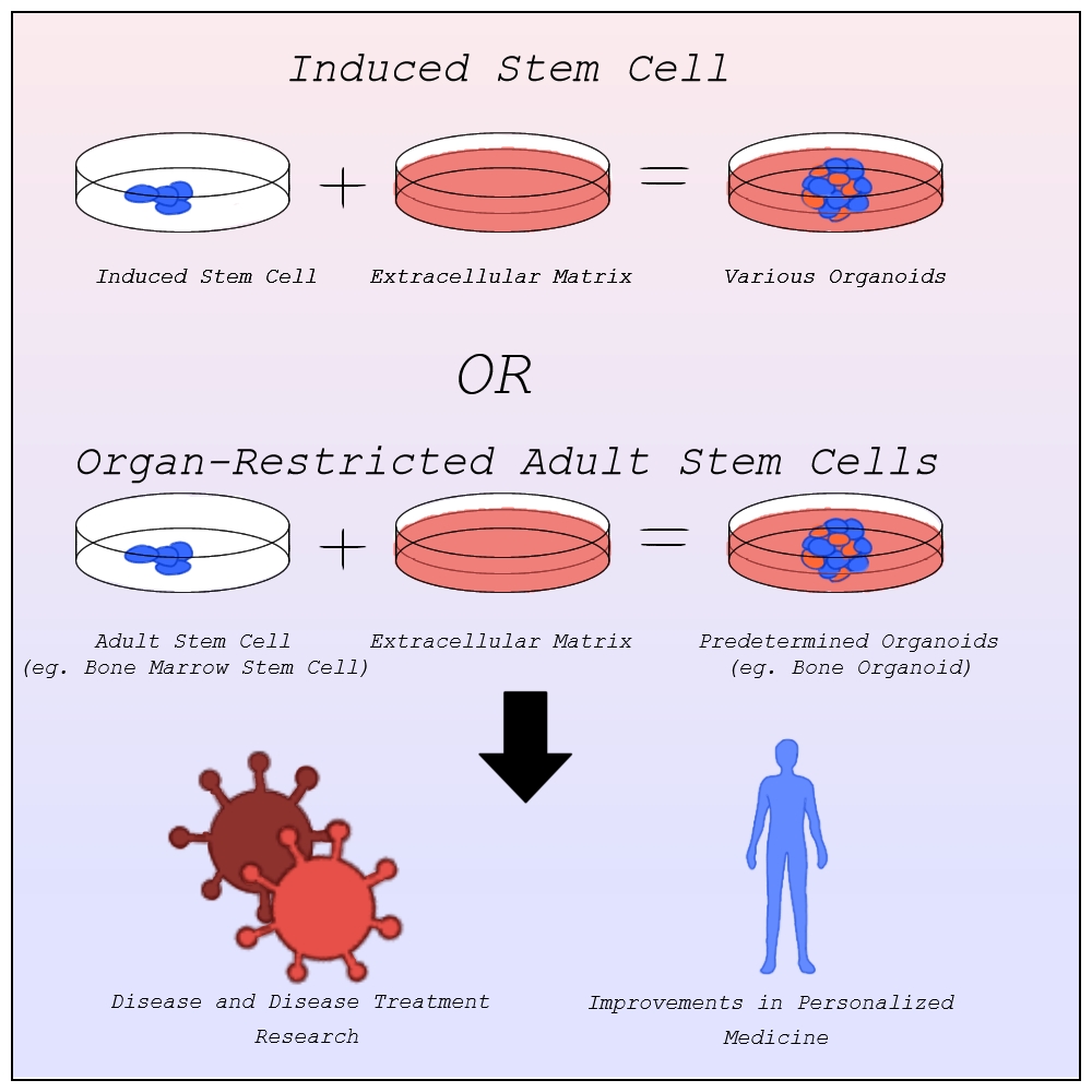
    Before the discovery of induced stem cells in 2006 by Dr. Shinya Yamanaka at Kyoto University, organoids were made from pluripotent stem cells as organ-restricted adult stem cells can be hard to access and are restricted to becoming cell types of their tissue of origin most commonly found in embryos (Agarwal et al., 2024). The line of when a zygote becomes a person, however, is blurry, raising many ethical concerns about the desecration of a person’s body. Induced stem cells allow for the change of specialized cells back into stem cells. Organoids can now be created from any tissue sample, entirely limiting the need for embryonic stem cells outside of studying early organ development (Frans Schutgens & Clevers, 2019).

    With better ways to develop organoids, scientists quickly realized their potential. Organoids offer unparalleled advancement in disease research and treatment, personalized medicine, and so much more that we simply don’t have time for here.

    Image by Alexandra Marshall

    Disease Research and Treatment
    One pivotal area of applications is understanding the development of diseases and disease treatment in humans. The lack of 3D structure in in vitro models lead to limited complexity and physiological relevance when studying diseases. Faulty predictions based on these studies can lead to treatments failing, with approximately 90% of clinical study drug candidates failing clinical trials and drug approval (Sun et al., 2022).

    Organoid models have none of these limitations. In 2013, the first human condition modeled using organoids, cystic fibrosis, was published. The model had “excellent predictive value” and became the first successful personalized treatment method for cystic fibrosis patients (Lancaster and Huch, 2019). The modeling of other diseases using organoids quickly followed from genetic diseases like Polycystic kidney disease and Alagille syndrome, to infectious diseases like Covid-19 and the Zika virus, to neurological diseases like Alzheimer’s and Parkinson’s, to multitudes of cancers, and more (Xu et al., 2018). The ability for organoids to be made of any human system means they can model any disease regardless of which systems they afflict.

    Organoids can be also grown co-culturally with bacteria allowing for bacterial infections and the diseases that arise from them to also be modeled and researched with organoids (Schutgens and Clevers, 2020). This has allowed for better understanding of bacteria like Helicobacter Pylori and Salmonella, aiding in detection and cures. As mentioned above, treatments can then be tested on these organoid systems without risking human lives or animal cruelty.

    Respiratory Syncytial Virus (RSV), a disease that is being modeled in airway organoids, causes around 66,000–199,000 deaths among children annually (Schutgens & Clevers, 2020). This is just one of the many diseases for which scientists are now better equipped to find viable treatments. Organoids have already saved millions of lives and their potential for future disease treatment has the potential to millions more.

    Personalized Medicine
    While these treatments work for many people, genetic and biological differences can impact treatment effectiveness and tolerance with some even leading to negative side effects (Breen, 2021). In such situations, personalized medicine that is specialized to fit these differences might be necessary. Personalized medicine, sometimes called precision medicine, challenges the idea that treatments that pass general trials will work for everyone, instead working to construct a treatment plan with a specific patient in mind best suited to their physiology.

    The idea of personalized medicine has been around for thousands of years, using one’s family’s history of diseases to diagnose the likelihood of that person having said disease. In the late 1900s the idea of evidence based medicine, the implementation of explicit reasoning based on a specific patient’s data, started being common practice when prescribing a patient (Visvikis-Siest et al., 2020). This opened the door for personalized medicine in modern science (Visvikis-Siest et al., 2020). This data was largely based on patients’ reactions to other treatments and family history however limited its effectiveness. The development of molecular biology and human genome mapping allows for a direct look at a patient’s physiology, aiding medical professionals in determining the biological reasons behind potential reactions to treatments before administration limiting adverse reactions.

    Organoids have greatly furthered this field. As mentioned above, organoids are made from a patient’s induced stem cells. This means that the organoid culture into a structure based on the patient’s DNA, containing all the same biomarkers and physiology that the patient would have at that level. Where other treatments could have varying effects from the tested population, organoids allow medical professionals to see the exact effects a treatment plan would have on the patient. The use of stem cells also allows organoids to grow much quicker than other models. This allows for quick and accurate testing of the effects of medication on a patient. One study published in Science involving gastrointestinal cancer therapies found that organoids would correctly detect when drugs would fail 100% of the time, and when the drug would succeed 88% percent of the time (Vlachogiannis et al., 2018). Once again, because of the diversity of systems that organoids can model, organoids can be used to model many different personalized medicine plans allowing for the variety of treatment testing needed to find the most optimal plan.

    Conclusion
    Organoids have had a monumental, yet widely unknown, impact on a wide variety of disease advancement and treatment developments and personalized medicine. These structures can then be used to model human tissues and organs with much higher accuracy and complexity than previously used non-human or in vitro models.

    Scientists use them to model disease development in humans and treatments designed for humans. This allows for a better understanding of the diseases and more effective treatments with fewer errors. They are also used to limit the risk of negative side effects from non-personalized medicine by furthering personalized medicine through the accuracy and speedy testing of many different treatment options.
    With continued and careful development of organoids, disease understanding and treatments, personalized medicine and so much more can continue to be improved, saving millions of lives.

    ————————————————————————————————-

    Work Cited:
    Agarwal, S., Saha, L., Sohani, A., Karri, U., Gor, R., & Ramalingam, S. (2024). The importance of niche in therapeutic strategies targeting normal and cancer stem cells. Cancer Stem Cells and Signaling Pathways, 15–33. https://doi.org/10.1016/b978-0-443-13212-4.00031-3

    Bredenoord, A. L., Clevers, H., & Knoblich, J. A. (2017). Human tissues in a dish: The research and ethical implications of organoid technology. Science, 355(6322). https://doi.org/10.1126/science.aaf9414

    Frans Schutgens, & Clevers, H. (2019). Human Organoids: Tools for Understanding Biology and Treating Diseases. Annual Review of Pathology Mechanisms of Disease, 15(1), 211–234. https://doi.org/10.1146/annurev-pathmechdis-012419-032611

    Generation and Cultivation of 3D Cell Models | ibidi. (2024). Ibidi. https://ibidi.com/content/1037-generation-and-cultivation-of-3d-cell-models

    Lancaster, M. A., & Huch, M. (2019). Disease modelling in human organoids. Disease Models & Mechanisms, 12(7). https://doi.org/10.1242/dmm.039347

    O’Connell, L., & Winter, D. C. (2019). Organoids: Past Learning and Future Directions. Stem Cells and Development, 29(5), 281–289.https://doi.org/10.1089/scd.2019.0227

    Schutgens, F., & Clevers, H. (2020). Human Organoids: Tools for Understanding Biology and Treating Diseases. Annual Review of Pathology: Mechanisms of Disease, 15(1), 211–234. https://doi.org/10.1146/annurev-pathmechdis-012419-032611

    ‌Simian, M., & Bissell, M. J. (2016). Organoids: A historical perspective of thinking in three dimensions. The Journal of Cell Biology, 216(1), 31–40. https://doi.org/10.1083/jcb.201610056

    Sun, D., Gao, W., Hu, H., & Zhou, S. (2022). Why 90% of clinical drug development fails and how to improve it? Acta Pharmaceutica Sinica B, 12(7), 3049–3062. https://doi.org/10.1016/j.apsb.2022.02.002

    Visvikis-Siest, S., Theodoridou, D., Kontoe, M.-S., Kumar, S., & Marschler, M. (2020). Milestones in Personalized Medicine: From the Ancient Time to Nowadays—the Provocation of COVID-19. Frontiers in Genetics, 11. https://doi.org/10.3389/fgene.2020.569175

    Vlachogiannis, G., Hedayat, S., Vatsiou, A., Jamin, Y., Fernández-Mateos, J., Khan, K., Lampis, A., Eason, K., Huntingford, I., Burke, R., Rata, M., Koh, D.-M., Tunariu, N., Collins, D., Hulkki-Wilson, S., Ragulan, C., Spiteri, I., Moorcraft, S. Y., Chau, I., & Rao, S. (2018). Patient-derived organoids model treatment response of metastatic gastrointestinal cancers. Science, 359(6378), 920–926. https://doi.org/10.1126/science.aao2774

    Xu, H., Jiao, Y., Qin, S., Zhao, W., Chu, Q., & Wu, K. (2018). Organoid technology in disease modelling, drug development, personalized treatment and regeneration medicine. Experimental Hematology and Oncology, 7(1). https://doi.org/10.1186/s40164-018-0122-9

  • Kidneys: The Innocent Bystanders

    When we purchase over-the-counter medicines, there’s often an assumption of their relative safety and harmlessness. After all, most ‘dangerous’ compounds are protected behind a prescription, right? Surely, if the directions on the back of the box or bottle are followed, no long-term health risks are possible, right?

    Unfortunately, this is often not the case. Long term use of NSAIDs — a class of common anti-inflammatory drugs including aspirin and ibuprofen — is associated with an increased risk of acute kidney failure.[1] Previously healthy young people have developed kidney problems due to long-term NSAID use while being none the wiser; kidney injury thus represents a generally unknown side-effect of NSAID use. Indeed, this feeds into a much larger idea within the field of healthcare — the role of the kidney as an ‘innocent bystander’. Often, acute and even chronic kidney injury are iatrogenic — an unintended byproduct of treatments for other diseases or conditions. NSAIDs aren’t the only common treatment that can negatively impact kidney health; diuretics are associated with an increased risk of acute kidney injury and even surgical procedures like heart transplants have been linked to acute kidney failure.[2,3] The susceptibility of the kidneys to iatrogenic conditions is often attributed to the high rate of blood flow through them and the resulting increased exposure to potentially nephrotoxic drugs.[4] After all, the kidneys are the blood filtration system meant to remove waste from the body; it makes sense that the drugs that enter the bloodstream and are filtered out may put a strain on the kidneys. Not just treatments, but even diagnostic techniques can have an adverse effect on the kidneys. The iodinated contrast drugs often used in diagnostic imaging have also been linked to acute kidney injury, with few suitable alternatives available.[5]

    With all these harmful pitfalls, preserving kidney health may seem like an impossibility when tackling complex conditions and comorbidities. However, awareness is a crucial first step towards improving patient outcomes. The kidneys are often put on the back burner when managing other conditions; physicians may prescribe NSAIDs, diuretics, and similar drugs that harm the kidneys over time while treating other conditions, or order diagnostic imaging that utilizes contrast fluid that puts a strain on the kidneys. On their own, these small actions may not have a significant effect, but it’s critical to be mindful of how treatments and conditions may intersect to cause renal dysfunction. By spreading awareness of the susceptibility of the kidneys to damage in these ways, individuals at higher risk for developing renal issues may be mindful about their use of certain over-the-counter medications and can develop a better toolkit with which to advocate for themselves.

    References:

    1 Dixit, M., Doan, T., Kirschner, R., & Dixit, N. (2010). Significant Acute Kidney Injury Due to Non-steroidal Anti-inflammatory Drugs: Inpatient Setting. Pharmaceuticals (Basel, Switzerland), 3(4), 1279–1285. https://doi.org/10.3390/ph3041279

    2 Ejaz AA, Mohandas R. Are diuretics harmful in the management of acute kidney injury? Curr Opin Nephrol Hypertens. 2014 Mar;23(2):155-60. doi: 10.1097/01.mnh.0000441150.17202.be. PMID: 24389731.

    3 F. J. Romeo et al., Acute Kidney Injury After Cardiac Transplantation: Foe or Common Innocent Bystander? Transplantation Proceedings 50, 1489-1495 (2018).

    4 Davidman M, Olson P, Kohen J, Leither T, Kjellstrand C. latrogenic Renal Disease. Arch Intern Med. 1991;151(9):1809–1812. doi:10.1001/archinte.1991.00400090095017

    5 Andreucci, M., Faga, T., Serra, R., De Sarro, G., & Michael, A. (2017). Update on the renal toxicity of iodinated contrast drugs used in clinical medicine. Drug, healthcare and patient safety, 9, 25–37. https://doi.org/10.2147/DHPS.S122207

  • A Spotlight on Black Licorice and its Darker Side

    Black licorice. You either love it or you hate it. Some delight in the intense spice flavor that comes with a bite of licorice and others find it repulsive and gag at the mere scent of it. As a divisive food and old-timey treat, black licorice has a long history of being a chewy confectionery and a popular flavor for drinks and tobacco in many parts of the globe. Licorice (scientifically referred to as Glycyrrhiza glabra) is a flowering plant that comes from the legume family. Its roots contain the flavor that we know as licorice and are commonly extracted for various uses. While it shares similar flavoring compounds, it is not related botanically to anise or fennel as some may think. (though sometimes they are used instead of licorice to evoke the same flavor profile)

    What makes black licorice so sweet?

    The iconic scent and flavor of licorice come from a mix of different compounds, but the one that gives it most of its sweetness is glycyrrhizin (a compound that is about 50 times sweeter than sugar!) This sweetness is different than regular sugar in that it more closely resembles sweeteners like Sweet ‘n’ Low which is comprised of saccharin. Some people vehemently dislike this type of sweetness which is also known to linger on the tastebuds for longer. Many people claim to dislike how “sickeningly sweet” it is and feel that it strongly dominates whatever you taste in your mouth for many minutes even after it is consumed. Others say that their aversion comes from the glycyrrhizin tasting more bitter than sweet.

    This bitterness can be attributed to herbal medicine, which licorice is also known to be useful for. There are many patented licorice-based medicines in China, where its extract is used in the treatment of bronchitis, colds, cough, and upper respiratory diseases. Glycyrrhizin, glycyrrhizinic acid, isoliquiritin, and glycyrrhizic acid are the most notable substances used for health benefits as they possess anti-cancer, anti-microbial, and anti-inflammatory properties. Excessive consumption that allows for the build-up of these compounds, however, may lead to serious adverse health effects.

    Consequences of Overconsumption

    Like most things in life, licorice is best enjoyed in moderation, but for more sinister reasons than you may think. Eating more than 57g of black licorice a day for at least 2 weeks could potentially cause serious health problems, such as an increase in blood pressure and an irregular heart rhythm (arrhythmia). This happens because of an excess of glycyrrhizic acid in the body. Normally your body is able to process glycyrrhizic acid into glycyrrhetinic acid, which is relatively harmless in small amounts, but when larger concentrations are present in the body, it can inhibit an enzyme that assists in converting active cortisol (the hormone that controls adrenaline) into inactive cortisone. You can imagine what too much active cortisol might do to your body, and this imbalance like this can wreak havoc on your body. 

    Cortisol is a hormone that plays a role in your fight-or-flight response and is similar chemically and structurally to aldosterone, which during a stressful event can signal to the body to hold on to water if you’re dehydrated. It does this by binding to structures in the kidneys that cause sodium to be pulled from urine in order to trigger osmosis and cause water to stay in your cells. Sodium has a +1 charge, however, and if it’s taken from the urine, then another +1 charge must replace it. This is done by potassium which can then replace the sodium in the urine and be excreted from the body. Now, when cortisol is present in concentrations that are dangerously high, it can be concluded that it would cause your body to pull more sodium from your urine, and you would lose more potassium right? 


    Cortisol (left) & Aldosterone (right)

    This leads to hypokalemia or low potassium presence in blood, which can be detrimental to the muscles of your body especially one of your most essential muscles, your heart. Potassium is an ion in your body that signals a relaxation of your muscles after a contraction. If there isn’t enough potassium present in your blood, then your muscles will stay contracted, which can be especially bad for your blood vessels and your heart. This is where the increased blood pressure and arrhythmia can restrict proper oxygen flow throughout your body and cause your organs to shut down.


    No Need to Panic

    Now, of course, the chances of this happening to the average black licorice fan are very slim as long as the treat is enjoyed in moderation. Since most people wouldn’t eat 57g of black licorice every day, I think it’s safe to say that fearing death by licorice overdose won’t be found in anyone’s nightmares. All in all, black licorice is a novel sweet with an interesting chemical makeup that is loved (or hated) by many across the globe.


    Sources:

    Pastorino G, Cornara L, Soares S, Rodrigues F, Oliveira MBPP. Liquorice (Glycyrrhiza glabra): A phytochemical and pharmacological review. Phytother Res. 2018 Dec;32(12):2323-2339. doi: 10.1002/ptr.6178. Epub 2018 Aug 17. PMID: 30117204; PMCID: PMC7167772.

    Wahab S, Annadurai S, Abullais SS, Das G, Ahmad W, Ahmad MF, Kandasamy G, Vasudevan R, Ali MS, Amir M. Glycyrrhiza glabra (Licorice): A Comprehensive Review on Its Phytochemistry, Biological Activities, Clinical Evidence and Toxicology. Plants (Basel). 2021 Dec 14;10(12):2751. doi: 10.3390/plants10122751. PMID: 34961221; PMCID: PMC8703329.

    https://www.healthline.com/nutrition/black-licorice-death

    https://www.youtube.com/watch?v=qg76qIZ4BQ8 

  • Racing Rats’ Impact on Mental Health

    A cartoon rat examines a rodent operated vehicle.Introduction

    The authors of “Enriched environment exposure accelerates rodent driving skills”, published in Behavioral Brain Research, conducted an experiment intended to study the impact of the environment on the efficacy of stress-releasing mental health treatments in lab rats. This study aimed to see how an enriched environment, both socially and intellectually, promotes stress relief while doing fun activities. The process for teaching the rats to drive the rodent operated vehicles (known as ROVs) also has implications for the importance of enrichment in how service dogs are trained.

    Study Summary

    There were two groups of rats that participated in this study. The control group of rats lived in a standard environment for laboratory rats, in standard laboratory cages, each with one other rat. The experimental group of rats lived in housing with an enriched environment. These rats got multiple rat friends, rat toys to keep them occupied, and multi-level housing.

    The study began with a “training period”, where rats in both groups were taught by operant conditioning how to drive the ROVs. While the authors of the study expected the rats living in standard housing to perform worse than the other rats, they were shocked at how much worse they performed. The authors noted that the control group rats failed to perform even “basic” tasks in driving the ROVs.

    After the training period (in which food rewards were used to encourage the rats to drive), both groups of rats continued to drive the ROVs even in the absence of rewards. However, rats living in enriched housing drove the ROVs without treats for a longer period of time than the rats living in standard housing. Throughout the study, the scientists measured the levels of hormones that were indicative of the rats’ ability to handle stress. Both groups showed an improved ability to handle stress as a result of ROV driving, but the enriched housing rats showed a significantly larger benefit than the standard housed rats.

    Conclusion

    This investigation shows that the environment of an animal has a significant impact on the effect of stress relieving interventions. The authors suspect that the enriched rats were more able to learn new behaviors (driving the ROVs) as a result of experiencing a wider variety of behaviors in their enriched home environments. The results of this study have interesting ramifications on the optimal conditions for other animals who will be undergoing rigorous training. Additionally, this study demonstrates how a good environment that has opportunities for social connection and mental challenges is beneficial in mental health treatment.

    References

    Crawford, L. E., Knouse, L. E., Kent, M., Vavra, D., Harding, O., LeServe, D., Fox, N., Hu, X., Li, P., Glory, C., & Lambert, K. G. (2020). Enriched environment exposure accelerates rodent driving skills. Behavioural Brain Research, 378, 112309. https://doi.org/10.1016/j.bbr.2019.112309

  • If I only had a…spine?

    For as long as I can remember, I have been obsessed with how people move. Now, hear me out. Even simple movements are fascinating if you really think about it. Electrical signals from your brain and spinal cord communicating with hundreds of muscles, forcing them to work together in a perfectly balanced symphony of contractions. All to maneuver our unwieldy skeletons gracefully through space.

    Do me a favor and stand up.

    For most of us, this movement feels like one of the simplest things we can do.

    Now, look at your legs.

    There are over 50 muscles below your hips alone. Yet, all these muscles just contracted in expert harmony to use the precise amount of force needed to move your body against gravity, all while maintaining near perfect balance. Precisely how we can perform these seemingly simple yet crucial movements on a whim is an active and exciting area of research, leading us toward innovation in movement rehabilitation, robotics, and beyond. These are movements we don’t even notice, like activating our muscles to breathe, blink, maintain our balance, or even walk. If you did notice these movements, you likely wouldn’t be able to focus on much else, transforming a simple grasp into an impossible and difficult task.

    Luckily, you don’t need your brain to do any of these things.

    More than just a cord

    Many people think of the spinal cord as just that, a cord. The cords and cables we typically interact with are charged with a very important but relatively simple task: bringing electricity from point a to point b. While the spinal cord is very important for bringing electrical signals from your brain to your muscles and organs, it does so much more.

    Picture an orchestra with a smart but rather lazy conductor performing for an audience. The musicians are like the motor neurons in the spinal cord, connecting to and contracting the muscles when the neurons fire, allowing you to move. Complicated musical pieces require guidance by our lazy conductor, just like throwing a dart or grasping an object requires guidance by the brain [1]–[4].  The audience’s cheers allow the orchestra to adapt, just like you use sensations from your body to improve or guide movements [5], [6].

    Yet, just like our experienced musicians don’t need the conductor to play simple or repetitive musical pieces, you don’t need your brain to perform “classic” movements. “The spinal cord is able to achieve so many behaviors by itself, completely isolated from the brain,” explains Dr. Cope, spinal cord neurobiology researcher and Georgia Institute of Technology professor.  “You can completely isolate the spinal cord in a living animal from the brain and it can walk on a treadmill [7], [8]. It can change speeds as the treadmill changes speed. You put an obstacle in its way, it can learn to lift its leg over that obstacle,” all without our lazy conductor [9].

    It was discovered in the early 1900’s that your motor neurons are fully capable of running the show [10]. “What that tells you is that there is this rich circuitry that in fact the whole motor system relies upon,” Dr. Cope explained. All in all, it looks like the spinal cord has the classics all worked out for you. Feel free to tell your conductor they can take the day off.

    How to run around like a chicken with its head cut off

    Unsurprisingly, experienced musicians are able to play complicated, intricate music without their conductor. Surprisingly to many, however, your spinal cord is able perform complicated, intricate behaviors without any input from the brain. How exactly are these behaviors possible? It’s all in the organization.

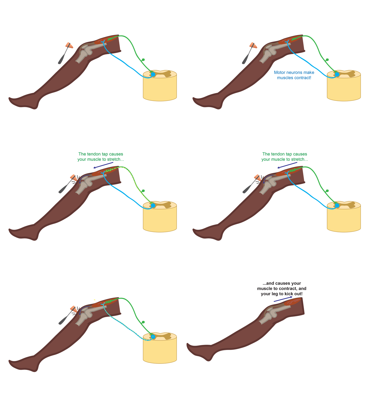
    If you’ve ever gotten a physical, you have probably experienced the odd sensation of your leg flying through the air without your consent. In the right spot, a simple tap on your knee by the doctor sends your foot on a trip automatically. We commonly refer to these types of movements as “reflexes,” in which no approval by the brain is required. This reflex pathway is relatively simple in organization; only two neurons are required, making this one of the fastest reflexes we have. One neuron senses the stretch of your muscle caused by the tap and immediately tells the second neuron to flex that same muscle. This flexion rapidly moves your leg before you can stop to think about it. This can happen in less than a few milliseconds [11]! You use your stretch reflex more often than your visits to the doctor, however. The stretch reflex helps you keep your balance without a second thought and is believed to be crucial for general sensory feedback and movement control.


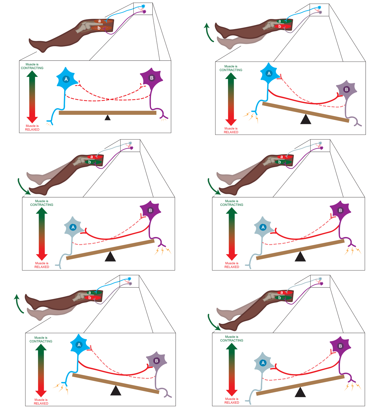
    What if we make things a little more complicated? Instead of just two neurons, let’s add in two sets of neurons in the spinal cord: set A controlling muscle a, and set B controlling muscle b. Much like a seesaw, these sets of neurons rhythmically alternate in activity. A neurons fire until they run out of juice, then B neurons take over and the cycle continues. With this small set of neurons, a pattern of alternating activity emerges. Together, A and B neurons form a central pattern generator. For humans, however, a 2-muscle central pattern generator isn’t very useful. Adding in more sets of neurons allows your spinal cord to rhythmically control more muscles in a more complicated pattern. With anything from breathing and scratching an itch to walking and running, the spinal cord is in charge.


    The patterns are there in your spinal cord, all you need to do is press start. “One of the things the brain does and can take full advantage of is to just send a ‘go’ signal to the spinal cord,” Dr. Cope explained. “[The brain] can say ‘Hey, all of the complicated things you do with timing and organizing … different muscles in different patterns, you do it. You’ve worked all that out. I don’t have to complicate my life with that.’” And while the brain can initiate and influence this pattern of alternating activity, it isn’t required. This pattern can just as easily be started by sensory input from your environment, or by sensory signals from throughout your body [9].

    At the end of the day, it seems like the spinal cord has it all figured out for us. But do we have the spinal cord all figured out? Not even close.

    The Mysterious Cord

    In the past year, the news has been abuzz with instances of paralyzed patients regaining the ability to walk [12]. Paralysis is typically caused by spinal cord damage. Up until recently it seems, spinal cord injuries often left patients with limbs that were difficult or impossible to move willingly, oftentimes without hope for improvement. So how are these patients taking these miraculous steps?

    A better question might be what happens to the spinal cord when it’s injured? We know some things about how it repairs itself, but we are far from the whole story. This means we are a far cry from fully repairing spinal cords ourselves. While these recent miraculous findings may make it seem like we have it all figured out, don’t let that fool you. “I think it’s exciting and I think it’s encouraging. I would say that we shouldn’t let our encouragement overshadow the fact that it’s nowhere close to what we want,” laments Dr. Cope.  “It’s going to require some basic neuroscience information about what the mechanisms are that are limiting recovery.”

    Researchers like Dr. Cope at Georgia Tech are working on a piece of this puzzle, studying to understand how the healthy and injured spinal cord contributes to and controls movement. Even with the great strides achieved recently by clinical studies [12], Dr. Cope explains that “We’re encouraged, but we have a long way to go.”

    References

    [1] D. P. Hanes and J. D. Schall, “Neural Control of Voluntary Movement Initiation,” Science (80-. )., vol. 274, no. 5286, pp. 427–430, 1996.

    [2] D. M. Wolpert and Z. Ghahramani, “Computational principles of movement neuroscience,” vol. 3, no. november, 2000.

    [3] D. A. Rosenbaum, K. M. Chapman, M. Weigelt, D. J. Weiss, and R. van der Wel, “Cognition, Action, and Object Manipulation,” Psychol. Bull., vol. 138, no. 5, pp. 924–946, 2013.

    [4] E. Freud, S. N. Macdonald, J. Chen, D. J. Quinlan, M. A. Goodale, and J. C. Culham, “Getting a grip on reality: Grasping movements directed to real objects and images rely on dissociable neural representations,” Cortex, vol. 98, pp. 34–48, 2018.

    [5] R. Shadmehr, M. A. Smith, and J. W. Krakauer, “Error Correction, Sensory Prediction, and Adaptation in Motor Control,” Annu. Rev. Neurosci., vol. 33, pp. 89–108, 2010.

    [6] D. M. Wolpert, Z. Ghahramani, and M. I. Jordan, “An internal model for sensorimotor integration,” Science (80-. )., vol. 269, no. 5232, pp. 1880–1882, 1995.

    [7] T. G. Brown, “The Intrinsic Factors in the Act o f Progression in the Mammal,” Proc. R. Soc. London. Ser. B, Contain. Pap. a Biol. character, vol. 84, no. 572, pp. 308–319, 1911.

    [8] S. Grillner and P. Zangger, “On the Central Generation of Locomotion in the Low Spinal Cat,” Exp. Brain Res., vol. 261, no. 34, pp. 241–261, 1979.

    [9] E. P. Zehr, “Neural Control of Rhythmic Human Movement : The Common Core Hypothesis,” Exerc. Sport. Sci. Rev., vol. 33, no. 1, pp. 54–60, 2005.

    [10] C. S. Sherrington, “Flexion‐reflex of the limb, crossed extension‐reflex, and reflex stepping and standing,” J. Physiol., vol. 40, no. 1–2, pp. 28–121, 1910.

    [11]  E. G. T. Liddell and C. S. Sherrington, “Reflexes in response to stretch (myotatic reflexes),” Proc. R. Soc. London. Ser. B, Contain. Pap. a Biol. Character, vol. 96, no. 675, pp. 212–242, 1924.

    [12] P. J. Grahn et al., “Enabling Task-Specific Volitional Motor Functions via Spinal Cord Neuromodulation in a Human With Paraplegia,” Mayo Clin. Proc., vol. 92, no. 4, pp. 544–554, 2017.

  • Batty or Bacterial?

    On The Origin of the Western Vampire

    The myth of a devilish creature draining the life force (blood) of a community was brought to life by European peasants. In the 1730s, this being was dubbed “vampire”, which came from the Slavic word “vampir”. The story of the vampire was spread throughout Europe by Austrian conquerors, who observed Eastern European protection rituals, such as removing the organs from corpses, and became swept up in the terror. Eventually, European immigrants carried the legends of the vampire to America.

    Bodily Basics

    Superstitions and mythology originate from some kernel of truth, some observable phenomenon. What were all these people seeing that made them believe an undead body was preying on the living in the night?

    Aspects of vampire mythology describe actual events that happen as a body decomposes. A corpse may appear to have “feasted” recently due to remaining liquid blood in the lungs being forced into the mouth. The appearance of a vampire victim as being “eaten” is typical of bloating that occurs during decay. The “screams” of a vampire is usually just gases escaping from the body. Additionally, bodies buried in colder areas may not appear decomposed at all, like the “living dead”. Taken together, it is easy to see why someone with no knowledge of the natural and scientific nature of these events would interpret them as supernatural.

    Contagion Explanation

    Vampire panics are not confined to a single spooky body. Over hundreds of years, small towns in America and Europe were terrorized by the idea that a vampire had come to turn family members and drain the community when in fact, these towns were being terrorized by outbreaks of contagious diseases.


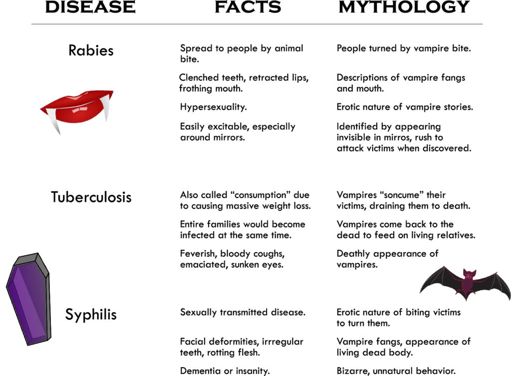
    A chart showing symptoms/effects of rabies, tuberculosis, and syphilis, and how these are similar to descriptions of vampires' appearance and behaviors.
    Similarities between some contagious diseases and common vampire mythology.


    Sources:

    Tucker, A. (2012, 10). THE GREAT NEW ENGLAND VAMPIRE PANIC. Smithsonian, 43, 58-65. Retrieved from http://prx.library.gatech.edu/login?url=https://search.proquest.com/docview/1086342252?accountid=11107

    Hammer, K. O. (2005). Vampires: The dark fantasy of the 18th and 19th centuries (Order No. 1449350). Available from ProQuest Dissertations & Theses A&I: Literature & Language; ProQuest Dissertations & Theses Global: Literature & Language. (305363212). Retrieved from http://prx.library.gatech.edu/login?url=https://search.proquest.com/docview/305363212?accountid=11107

    Burcum, J., & Writer, S. (1998, Oct 28). Putting the undead to rest // researchers have tried to shed light on the vampire myth, saying the horrific blood feasters were merely the victims of rabies, blood disorders or superstitious imaginations. Star Tribune Retrieved from http://prx.library.gatech.edu/login?url=https://search.proquest.com/docview/427048134?accountid=11107

  • Varicella-Zoster Strikes Again

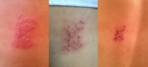
    Shingles on day 3, 6, and 11
    Shingles on day 3, 6, and 11

    I had chicken pox when I was 8 years old. This was before there was a vaccine for varicella zoster, the virus that causes chicken pox. When I was growing up everyone got the virus and it made you feel like crap and gave you sores all over (most people wound up with at least a few scars) and then you got better. In fact, when the vaccine was introduced in 1995 many people thought it wasn’t necessary because the disease was relatively mild. The 10,000 people hospitalized each year in the US because of complications and 100 people who died every year probably didn’t agree.

    And you know what? I don’t agree either. Varicella or Herpes zoster is a tricky virus that goes latent in nerve cells, much like its cousin Herpes simplex virus. Basically this means that when you recover from your infection, some virus hides out, waiting for your immune system to get weak from stress, old age, other infections, or whatever and then it attacks again! I don’t know what triggered it in my case, but a few days ago I got shingles. Shingles is a yucky, sometimes terribly painful, outbreak of the same virus that causes chicken pox. This time I didn’t get a fever and sores all over, just a cluster of sores along one of the nerves on my back. The scary thing about this, though, is that I am infectious. I could actually give chicken pox to someone who hasn’t had a chance to be vaccinated (a newborn baby for example), a pregnant woman, or anyone who is immunocompromised for some other reason like HIV disease or because they’ve had a transplant.

    I could also give chicken pox to people whose parents chose not to vaccinate them. These could be children or adults. Unfortunately, they didn’t get a chance to decide whether they wanted to get a life threatening illness because their parents thought it would be “healthier” than getting a vaccine. They also won’t get to choose when they get shingles, and will put other completely helpless populations at risk when they get infected.

    Some anti-vaccine people perpetuate the myth that being infected and recovering from a natural infection provides better immunity than getting a vaccine. This is simply not true. Viruses have evolved sly ways of manipulating our immune system, and often the immunity you develop from natural infections will not protect you very well from being reinfected or from the reactivation of latent viruses.

    You know, if I had been born a decade later, I would have been able to get the chicken pox vaccine and I wouldn’t have this icky rash on my back. If I were older, my doctor would have recommended the shingles vaccine for me.  Unfortunately I am stuck in the middle and got sick, but you don’t have to.  Go get the varicella vaccine, and get your children vaccinated.

  • Badass Biology: Toxoplasma gondii

    Toxopic

    Look, we humans are awesome. We own this planet. If we really wanted to, we could spill every barrel of oil we have into the ocean, light it all into one gigantic fiery blaze, get on fire-proof boats in robot suits, somehow put on war paint inside the robot suits, build obnoxiously large metallic middle fingers, and shout to the heavens “TRY TO IGNORE THIS, MOTHER EARTH!!” Then, if we try really really really hard, we could clean it all up again, because some of us are actually sorry and some of us like to keep our marine food webs and all.

    Why orchestrate such a display of power? Because we are the all-powerful movers of earth, the masters of fire and brimstone, and the makers of Haribo Gold Bears because holy freaking balls those things are really freaking delicious. We also have days of the year set aside where we butcher and devour different animals in front of children, purely because that’s what we’ve done for centuries. That right there is true power.

    Point is, we are masters of everything we see. Remember in the Lion King where Simba is sitting with Darth Vader, who I guess came back to life to inhabit the form of a lion, and Vader is all “everything the light touches is our kingdom”? Yeah, we humans are exactly that. The only difference between humanity and Darth Vader is this: while the lions behave at the whim of the team of animators and writers that brought them to movie stardom, we are being toyed with by a tiny microscopic parasite that enter our guts, hitchhike a one-way ticket to our brains, and deliberately try to kill us by making us do really stupid and risky things.

    I bet if you were eating a sandwich right before that last sentence, you probably aren’t anymore. It’s hard to eat something that’s been catapulted by your own shocked reaction, sending sandwich chunks to the opposite wall mid-chew. Even then, the walls may be really dirty, and that’s just not sanitary. I have no idea when I washed my walls last, and I keep my room relatively clean.

    Why is this parasite trying to kill us? Simple. They want to get to our cats.

    DAMMIT, MR. SCRUFFLES!!!

    One of the reasons I wanted to write this article was to make myself look amazing. This isn’t hard, considering my creative wit and depth of scientific knowledge is nearly endless. The main reason I wanted to write this, though, was to talk about some badass ways that parasites can take over the minds of their hosts. It’s actually not uncommon to find mind-controlling parasites in nature. In fact, there is an entire field, called neuroparasitology, focusing on the effects a parasite can exert on host biochemistry, neurophysiology, and ultimately behavior and ecology.

    Science jargon aside, there are some pretty badass parasites out there that tend to turn hosts into zombies. For instance, a genus of fungus called Chordyceps  invades the mind of its insect host, forces it to climb to a high place, orders its newly-induced slave to latch on with a deathly grip, and busts out of the host all Alien-styled to shower its lethal spores onto unsuspecting insect hosts below.

    Huuuugggghshhh, gross. I’m sorry I did that to you. I even feel a little dirty describing that. So, in an effort to make you and me feel less creeped-out and anxious, I’m going to comfort you by giving you increasingly creepy and troubling examples.

    So there’s this non-sarcastically beautiful creature called the jewel wasp that wrestles cockroaches into submission and bites them in the neck to inject a mild dose of venom. This venom paralyzes the roach, and after a short time, causes the roach to move with absolutely no will of its own. The wasp then literally starts a one-float cockroach parade, leading the roach to the wasp’s burrow like a Siren leads a ship’s crew to the abyss to be eaten by mermaids or something. Gotta admit, I don’t fully understand the Sirens’ intentions. The wasp seals the roach inside the burrow with a single egg and a comically large timer counting down the exact moment until the egg hatches. When the timer goes off, the cockroach is devoured to death by the larvae and pupae of the wasp, giving rise to another generation of the most beautiful thing in the universe made entirely of cockroaches. Technically, these kinds of parasites are called parasitoids because they live on or within the organism for a long while, but they ultimately lead to the sterilization or death of their hosts.

    The jewel wasp: made from 100% real cockroaches!! Holy balls, I just shuddered. Like, that seriously just happened.

    Another badass parasitoid wasp, called Glyptapanteles, lays its eggs in a caterpillar to develop and consume the host. This time, however, the host does not die. IT DOES NOT DIE. No, these tenacious caterpillar hosts, half-devoured and all, cling to life like that chip I just dropped into my bowl of homemade salsa.

    I hate when this happens.

    However, the wasp is not yet finished with its host. The caterpillar, after having live wasp pupae burst from its insides, spins a web of silk over the developing cocoons, stands upright on its back legs, puts on a lucky Japanese headband, and guards the cocoons with its life. Seriously, I wouldn’t dare screw with a half-eaten caterpillar sentinel, not even if I got a tummy full of wasp bodies as a treat afterwards. Eventually, the caterpillar starves to death, and that sucks, but at least– OK seriously curtains, what the hell are you doing?! Get it together!!!

    OK, so I just bought one curtain yesterday, and not only is the color subtly different than the other curtains in my room, but they’re about a foot shorter, too. It keeps moving and I keep staring at it thinking “I can’t wait to give you back to the store”. I have never hated a curtain so much in my entire life. Please, curtain, quit moving and let me write.

    Anyway, the point is that there are some pretty bitchin’ parasites and parasitoids that turn their hosts into zombies. You’ll notice though that all of the host examples I’ve used so far are from insects. Insects are little and dumb, and their tiny dumb stupid heads are really easy to invade. I mean, we’ve got huge brains! HUUUGGEEE!!! And they’re so complex, too! I mean damn, your visual cortex is so complicated that if you took every grain of sand on a 50-meter stretch of beach beach and lined them end to end, I’d be really disappointed because you decided to do that instead of listen to me talk about the visual cortex.

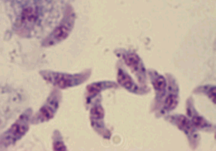
    No, we are not immune. You read the first couple of paragraphs, so I assume you knew that already. So let’s just jump right into things and talk about Toxoplasma. This little protozoan is microscopic, but somehow it’s able to hijack the most complex brain on the planet like it ain’t no thang.

    Braaaaainnnssss. Rat Amygdalaaaaaaaasssss.

    Much like a forgotten old woman with nothing to live for, Toxoplasma loves cats. I’m not sure why Toxo loves cats so much, but in reality this is not very strange. See, parasites often have to infect several hosts in order to reach the one it’s really out to infect, called a primary host. All the hosts it infects in order to reach the primary hosts are called secondary hosts, or intermediate hosts. Secondary hosts often don’t feel the full effects of infection because the parasite isn’t reproducing inside of them, but occasionally a secondary host can have its behavior manipulated in order to reach the primary host. This is what Toxoplasma does. In fact, Toxo does this very very well.

    The most clear-cut example of Toxo behavioral hijacking comes from the changes it can make to the brains of rats. Since Toxo is out to infect cats, it often causes the prey of cats to become more apparent in hopes that its current host will end up in the cat’s belly, the perfect place to make little Toxo babies. How does it cause rats to become more apparent? It forms cysts in the rat’s brain. In particular, Toxo cysts form near a region called the amygdala, a tiny region that controls fear and aggression. These inhibit the function of the amygdala, and basically cause rats to get sexually turned on by cat odors and actively seek cats by tracking their urine. Seriously not kidding.

    Toxo gets really really interesting when we start talking about humans. The tiny protozoan can cause the formation of brain cysts just as it does in rats, though the damage is very subtle at first. These cysts aren’t as selective in their real estate options, so they just kinda form wherever. They release enzymes that convert a dopamine precursor into dopamine itself, triggering the action of receptors of a group of neurotransmitters called catecholamines (which include dopamine, epinephrine, and norepinephrine). Ultimately, this causes people to do some wonky things.

    What kind of wonky things? Well, for one, do you remember the Beatles?  Well, John Lennon once saw a newspaper article about a car crash and wrote a song about it, which isn’t really relevant but does serve as a good segue into talking about how Toxo can make you crash your car. Just as in rats, Toxo can induce reckless behavior. So basically…man, I gave it away. Gimme a second to recover.

    Ah, screw it. Here’s an article on how car accidents are caused mostly by Toxo victims.

    What else can Toxo do, you ask?? Well, it’ll deform your babies, of course!! Apparently Toxo can cause some pretty terrible damage to babies while they’re in the womb, causing deformities that I cannot describe to you. Let’s just say that I googled “Toxoplamodium babies” and it’s not likely that I’ll sleep tonight because of that decision. You’re going to google it anyway, aren’t you?? You heartless bastard.

    See? It’s a real problem because this floating baby head appears on a banner with the name of the disease. That means it’s dangerous.

    These things all seem pretty obvious. Sure, Toxo can cause people to be reckless, and babies are easy to injure as it (Anderson and May, 1967) is so the damage from Toxo seems a bit expected. What else can Toxo do that’s more unexpected? To find out, let’s take a quiz.

    Question: Which of the following facts about Toxo is true?

    A)     Toxo can cause adult humans to become incredibly attached to their cats

    B)      Toxo can spread through dog semen, and can help upregulate testosterone to increase the odds of infecting a female

    C)      You’ll have no symptoms of Toxo in the later stages of infection

    D)     Toxo is associated with causing schizophrenia.

    Answer: all of the above.

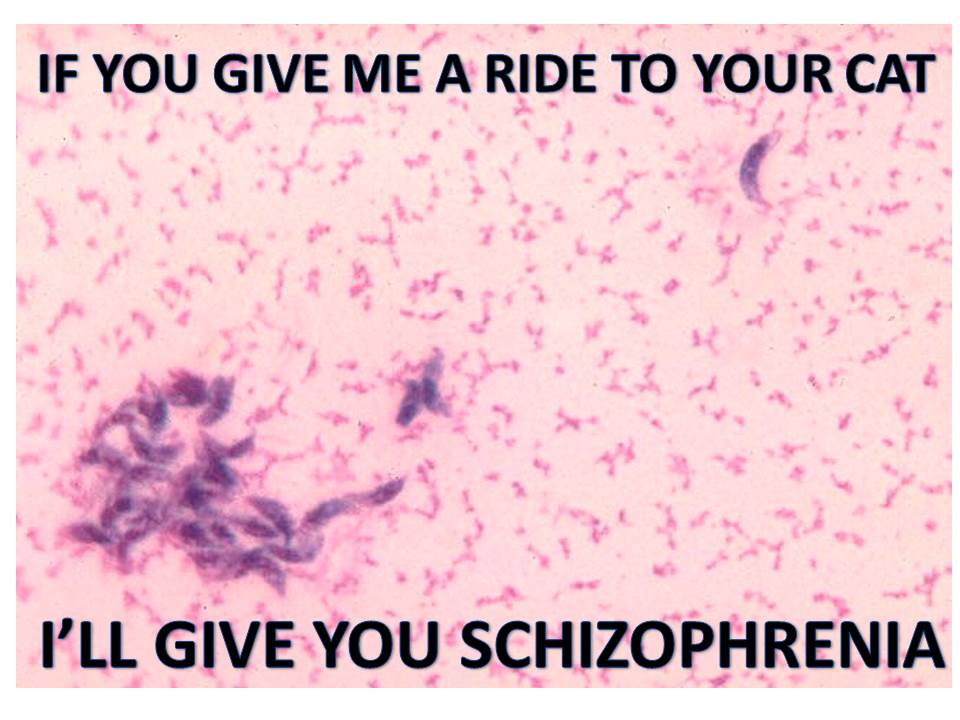
    I find it absolutely mind-blowing that a tiny protozoan can increase a human host’s risk of schizophrenia. This isn’t some brain augmentation to an ancient area like the amygdala. Inducing hallucinations is not simple to do. On top of that, Toxo can cause humans to acquire ADD, OCD, and can even increase suicide risk. Just read this link. Your mind will be blown too.

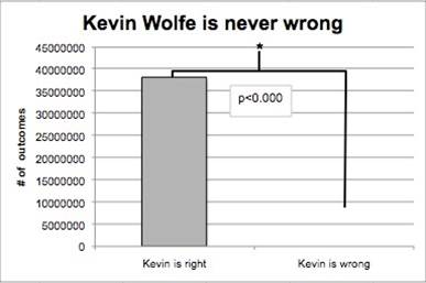
    Nature is truly out to kill us. How do I know? I am never wrong.

    Dat p-value.
    Dat p-value.

    How else do I know? Toxo exists. We think we’re so high and mighty, immune from crazy zombie-inducing parasites.We are clearly not. Here’s where I make a point that we are not so different from components of the greater aggregate of beings we call “nature”, and that we ourselves are indeed a part of that. Then, here’s where I get really preachy, talking about how things we do affect the natural world, and here I mention the circle of life and make a funny quip about the Lion King and the irony therein that my argument of the circle of life has referenced an earlier part of this article.

    Want to hear me talk about an animal, plant, bacterium, or whatever the hell else that you think is awesome? Leave me a message in the comments! I’ll probably ignore it, unless it’s actually really good. It’s worth a shot to try though.

    Come on. Comment. Go for it.


    To view the really interesting article most of this information came from, click this link!

    Photo credits:

    -http://listvisit.com/wp-content/uploads/2012/12/grey-american-shorthair-cat-3.jpg

    -http://pcdn.500px.net/7757320/7090f93b289604fab91e62214d1243b8f13ae184/4.jpg

    -http://media.tumblr.com/tumblr_lfp2sreXdv1qc9f5v.gif

    -http://kidshealth.org/parent/infections/parasitic/headers_92320/P_infectionB_toxoplas_1.jpg

  • Bizzare Medical Treatments: Bayer Heroin

    Bayer heroin advertisement.

    Bayer AG, a multibillion dollar, German pharmaceutical company is regarded as one of the leaders in its sector, producing mild painkillers such as Bayer Aspirin and Aleve, the vitamin C supplements Redoxon and Berocca, and many other well known drugs. This reputation combined with the angelic sounding tagline on their website, “Bayer: Science For A Better Life” makes it hard to believe that in 1895, the company marketed heroin as a cough suppressant.

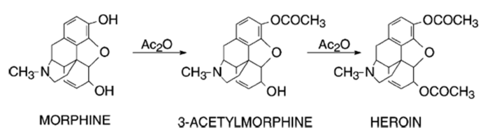
    Acetylation of morphine, by way of acetic anhydride (Ac2O), to synthesize heroin.

    Heroin is an opiate synthesized by adding two acetyl groups to morphine, found in the opium poppy. When Bayer introduced this new drug, it was actually meant to be a non-addictive substitute for morphine, a common cough suppressant back then. Little did they know that their well intentioned discovery would eventually become one of the most abused drugs, known for its destructive effects on the human body. It turns out that heroin crosses through the blood brain barrier 100 times faster than morphine because the acetyl groups makes it highly soluble in lipids; once it crosses the barrier, it actually turns back into morphine. Essentially, heroin is a prodrug, a class of inactive drugs that are converted into active form in the body by normal metabolic processes. To the Bayer scientists, heroin may have looked less potent from morphine on the lab bench, but in the body, it ended up being a faster acting form of morphine, making it extremely dangerous and addictive. Needless to say, even though heroin actually does help stifle coughs, Bayer eventually pulled this product off of their catalog.

    Though today, the possession or selling of heroin is illegal in most parts of the world, it can actually be used in the United Kingdom legally, under careful control,  as an analgesic and a maintenance drug.


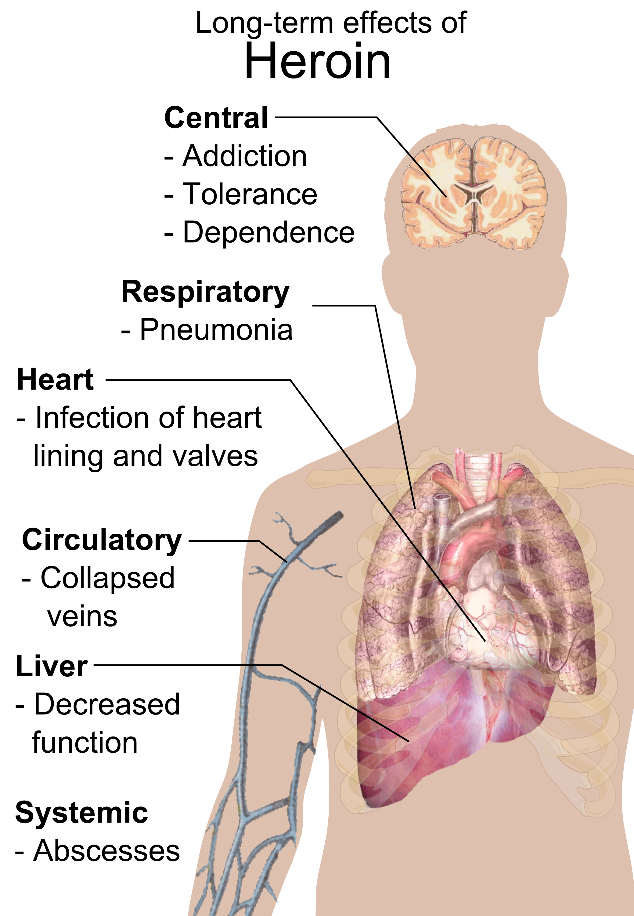
    Common long term effects of heroin.


    Image Sources:

    http://en.wikipedia.org/wiki/File:Bayer_Heroin_bottle.jpg

    http://en.wikipedia.org/wiki/File:Long-term_effects_of_heroin.png

    http://www.opioids.com/heroin/bayerheroin.jpg

    http://optiond.wikispaces.com/file/view/heroin_synthesis.png/116773659/711×189/heroin_synthesis.png