NOTE: This post is not related in any way to the 1956 sci-fi film that depicts an extraterrestrial invasion where the invaders “steal” human bodies. This is NOT a review of the Invasion of the Body Snatchers (though I do highly recommend the film).
Rather, this is brief overview of a period when robbing bodies, often from graves, was used as a means to provide physicians with subjects for dissections.
When?
Body snatching was most prevalent during the seventeenth to nineteenth century, though there have been reports of this activity occurring as early as the fourteenth century.
Why body snatching?
– Surgical practice (High Demand): Most fifteenth century physicians scorned and considered it degrading to work with their hands; They often stood by patients they treated and ordered servants to perform what they thought should be done by hand. This disconnect between the physician and surgery created a void in anatomical and surgical knowledge. During the sixteenth century, Andreas Vesalius, a Flemish physician, led a movement to fill that gap. His efforts culminated in the publication of De humani corporis fabrica, one of the greatest anatomical textbooks. During that time, Vesalius traveled around Europe performing dissections himself, generating interest in surgery and leading physicians to embrace the values of a hands-on approach to medicine. This surge in interest in surgical practice created a great demand for bodies to be used for dissection.
– Shortage of bodies (low supply): Two centuries ago most states did not permit the use of unclaimed bodies for dissections and most of the states that did limited them to executed criminals. However, this was not enough as executions were few in the states. For example, Massachusetts executed less than 40 persons from 1800-1830. In addition, as the number of medical schools increased, so did the demand for bodies. This legal blockade created a shortage of bodies available for medical schools forcing them to turn elsewhere.
–Body decay (high turnover): Lack of refrigeration was a common problem back then; not only did it spoil food, but it spoiled bodies. This meant that there was a high turnover rate; physicians had a limited time frame to dissect and study bodies after death. This created a situation where there was a constant demand for bodies.
SOLUTION: Where can you find a large supply of bodies? Graveyards.
Who stole bodies?
-At first, grave robbers were medical professionals, students, and hospital porters who stole bodies for their own purposes. However, as time passed, medical schools advised students, faculty, and personnel to avoid engaging directly in stealing bodies. From there grew a class of individuals known as resurrectionists or body snatchers, men who made a living exhuming corpses and selling them to schools. Eventually, body snatching organizations/gangs started to appear. As laws were put in place, robbing became more profitable; their task became increasingly difficult and dangerous, and the price of their commodity increased.
-Since body snatching was an arduous task, it was often men who participated in this activity, though women were sometimes involved in non-physical tasks or snatching bodies not yet buried (falsely claiming a body to be buried).
Whose body was stolen?
-Race, sex, and social status did not matter. Nobody was safe from disinterment.
Note: Though records show that people of all race, gender, and social status were victims, the golden era of body snatching coincided with the period of racial and social inequality in the states. This meant that immigrants, blacks, and the poor were most often targeted.
The Art of Body Snatching:
Many considered body snatching an art. Some texts describe the act in lengthy detail spanning several pages, detailing tactics on how to procure bodies as efficiently as possible. I will spare you from the lengthy details without skipping over the essential techniques.
Step 1: Secure knowledge of a prospective burial: Information of a burial was usually supplied by mail or messenger. Such a message was often sent in code and not directly to the resurrectionists, but through some intermediary, such as a druggist.
Step 2: Scope the location: The grave should be accurately located during daylight hours so that a search in the dark would be unnecessary. A common method was to reconnoiter as a hunter. A stranger would appear in the neighborhood on the day of burial with a shotgun over his shoulder, apparently hunting small game.
Step 3: Wait until nighttime
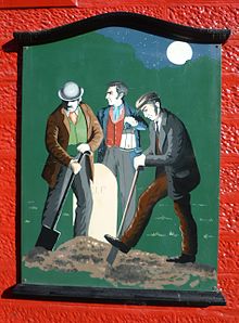
Step 4: Begin the disinterment
- Plan: At least two men and a watcher/driver were needed in a disinterment. One man remained with the transport; that person would drop off the other three at the location and return at a specified time. The other two removed the body.
- Execute: The process of disinterment was fairly straightforward: a hole would be dug directly above the coffin, the coffin would be opened, and finally, the body would be pulled out. Since graves were dug to be about 5 ft deep (from the top of the coffin), extracting a body could be quite laborious. To make the extraction easier, resurrectionists used a modified pole to drag bodies out of graves. This pole was hooked at one end and had grips at the other (similar to the letter ‘J’). The hook would be placed under the chin of the deceased and the men would push or pull the body out using the other end of the pole. As you can imagine, this ‘hooking’ technique often caused damage to the corpse.
Step 5: Clean Up: The grave should look as it did before the disinterment to avoid suspicion and capture.
Ethics
The seventeenth to nineteenth century was a great era for medicine. Discoveries in anatomy led to improved surgical treatments and healthcare. However, people often forget that many of these advancements came at the expense of bodies being robbed. Families would go to cemeteries to pay respects for loved ones only to find the grave(s) in disarray, or would pay respects without knowing the remains of their loved were gone. Until the adoption of more coherent and stricter laws regarding body donation, many physicians considered body snatching a necessary evil.
__________________________________________________
Sources
Nuland, Sherwin B., Doctors: The Biography of Medicine. New York: Vintage, 1995. Print.
Waite FC. 1945. Grave robbing in New England. Bull Med Libr Assoc, 33:272–294.
Humphrey DC. 1973. Dissection and discrimination, the social origins of cadavers in America 1760-1915. Bull NY Acad Med, 49:819–827.
Frank JB. 1976. Body Snatching: a grave medical problem. Yale J Biol Med, 49:399-410
Regis Olry. 1999. Body Snatchers: the Hidden Side of the History of Anatomy. J Int Soc Plastination. 14(2): 6-9
Image:
http://en.wikipedia.org/wiki/File:Body_snatchers_at_work,_Old_Crown_Inn,_Penicuik.JPG