Category: Articles

  • Organoids Part Two: Despite Limitations, They’re Still Amazing

    Clearly one article wasn’t enough to explore the full potential of organoids, so here we go again. This article won’t make much sense without reading the first but a quick catch up:
    Organoids are 3D cell structures grown from Stem cell samples and grown in nutrient rich gel.

    • They can better model human systems as they are more complex than their 2D counterparts and a better representation of human systems than animal models.
    • This advantage makes organoids uniquely beneficial to the field of drug research and testing and personalized medicine.
    • Link to original article if desired: https://2026.chargedmag.org/2026/04/organoids-what-are-they-and-why-theyre-amazing/

    Limitations
    These applications are not without limitations however. While organoids can replicate many aspects of human tissue, some specific cell genes are harder to find in organoids (Andrews and Kriegstein, 2022). This impaired the organoids’ ability to properly simulate the systems they are derived from, preventing researchers from properly modeling diseases and treatments in that area.

    Additionally, while developments have been made to improve organoid lifespans, organoids still require controlled environments and regular care to survive (Simian and Bissell, 2016). Even the longest lasting organoids, neurological models, have only survived a couple of years. Many organoids will even grow patches of dead tissue in the center due to a lack of oxygen hindering their research capabilities (Andrews & Kriegstein, 2022). In organic systems, cells are constantly interchanging molecules to maintain their functions and prevent necrosis. The nutritional gel can only do so much to get the molecules to cells at the interior of organoid structure without the natural systems to supply it.

    All of this causes organoid maintenance to be costly compared to in-vitro models, which are relatively cheap and easy to handle (Schutgens & Clevers, 2020). This stifles research leading to many knowledge gaps in the field. Furthermore, these restrictions limit modeling larger systems. Potential treatments tested in smaller organoids could have unpredictable impacts on a larger system when prescribed. Concerns around this limits applications of organoids, creating Theory-Application Voids.

    There are also many population gaps in organoid research as certain demographics are underrepresented in the stem cells used to create the organoids. These stem cells are often pulled from stem cell donor registries which have significant demographic, systemic, and behavioral biases. Further development into organoids and increased diversity in biobanks can increase organoid capabilities and applications.

    Concerns
    There are also current concerns surrounding organoid research that need to be addressed. While organoid models no longer rely on stem cells taken from embryos, they are still used in organoids modeling traditional embryo frameworks. These models use STEM cells to study the process of early embryonic development and can be very useful in identifying gestation concerns or potential risks to a fetus (Pereira et al., 2020). Once again, the line of when a zygote becomes a person however, gets blurry. Multiple public figures and many private citizens are debating if the creation of an embryo, even as a limited organoid, for study is ethical.

    There are also concerns about neurological organoid models used for neurological diseases (Lavazza and Massimini, 2018). Current models can only imitate parts of the human brain. However, as models more closely resemble a human brain with increased functionality, ethical questions surrounding the creation and experimentation of something than can think and feel will only grow. The line being living, sentient, and sapient is hard to discern, especially with limited structures that might not have the ability to communicate mental facilities.

    Future implementations of organoid models also raise ethical concerns regarding their financial exploitation and patient consent. People’s cell matter must be collected and added to databases to be used in organoid research (Bredenoord et al., 2017). Active patient involvement in organoid development is essential when considering this process. Their participation in cell donation must remain voluntary and properly compensated, and this is currently not guaranteed.

    The implementation of organoids in personalized medicine could also have complications on the healthcare system (Stefanicka-Wojtas & Kurpas, 2023). While organoids could improve personalized medicine, the increased specificity of treatments could lead to decreased procedure lifespans and certainty. The underrepresentation and price of organoid backed research could also exacerbate existing disparities in healthcare and lead to an increase in general healthcare costs or dropped plans due to cost-shifting (What’s Behind Rising Health Insurance Costs? | Johns Hopkins Bloomberg School of Public Health, 2025).

    Scientists in organoid research have repeatedly advocated for remaining cautious and careful in this relatively unexplored ethical field.

    Potential Applications
    Despite all of that dour news, organoids are still having a positive impact on millions of people with the potential to aid millions more. Unfortunately we only have time for two of these here: Regenerative Medicine and Transplantation.

    Regenerative Medicine is the field of science dedicated to the regeneration and restoration of human tissue based in Stem cells. Many different animals have regenerative properties from the regrowth and axolotl limbs to sea cucumbers regrowing internal organs to flatworms regrowing entire heads. Throughout history scientists have been inspired by these properties and researched the mechanisms behind them, leading to the field of regenerative medicine. While the study of artificial organs has been around since the 1930s, the modern idea of regenerative medicine centered around Stem cells started in the 1990s (Sampogna et al., 2015).

    As mentioned in my first article, the very purpose of Stem cells is to replace damaged, specialized cells. By comprehending the properties behind their functions, scientists can apply them to other areas. It is organoids that allow scientists to grow the cell cultures and tissue samples that are behind regenerative research. If someone has a large wound or damaged organ, an organoid of that structure could be applied and integrated into the person’s natural system. The organoid could be made from the person’s own cells, bypassing an immune response that comes with many non-biomaterials or transplants. This has already been proven promising. In one study, organoids made from the stem cells of mice suffering from inflammation and damage of their colon lining were successfully transferred onto the mice’s injured tissue. The cultured tissue covered the damage leading to a decrease in inflammation and harmful symptoms (Babak Arjmand et al., 2022). There are currently limitations, such as the delivery of nutrients mentioned above that directly challenge this field, but with more research scientists can find better ways for organoids to improve people’s lives through regenerative medicine.

    Transplantation is a great example of the applications of regenerative medicine. As mentioned above, tissues and cell cultures can be grown using organoids. This is not the extent of their potential however. As organoid research develops, scientists are working to make larger and more complex models. Through assembloids, the combinations of different types of organoid (a topic for a later article), lab made organs or entire organ systems could be created from a single stem cell. In 2025, scientists were able to successfully transplant pig kidneys made from organoids (SMC Spain, 2025). In the future organoids could be used to transplant an entire human heart or cardiovascular system. An even farther yet still plausible possibility would be the creation of entire limbs for transplants with scientists already researching limb organoids (Tsutsumi & Mototsugu Eiraku, 2023). Approximately 5,600 people in the U.S. die each year simply waiting for a transplant in addition to the 30% of recipients who die within 10 years of a transplant due to complications such as transplant rejection (What Are the Complications of Transplantation?, 2019). Organoids have the potential to prevent these deaths, and while there are still complications due to the price and longevity of organoids, they will only improve with continued exploration.

    Conclusion
    Organoids are amazing. They have the ability to impact almost every field of biomedical science and save millions of lives. While this article does take a much more nuanced look at organoids, diving into their limitations and complications, scientists are aware of these issues and taking careful approaches to research. Improvements in organoids, especially to their limitations, can only be made if research is supported and continued. There is still so much potential for organoids that we have only just scratched the surface of and so many more applications that we haven’t even considered yet. Organoids have even been proven to work with many other biomedical instruments and tools, aiding in their development as well. The possibilities are endless.

    Organoids are my passion, I’ve been interested in them for years now. Yet no one knew what I was going on and on about. I hope that these articles inspire curiosity and spread the wonders of organoids to others.

    ————————————————————————————————

    Works Cited:

    Title Image: Illustration created by Alexandra Marshall

    Andrews, M. G., & Kriegstein, A. R. (2022). Challenges of Organoid Research. Annual Review of Neuroscience, 45(1), 23–39. https://doi.org/10.1146/annurev-neuro-111020-090812

    Babak Arjmand, Rabbani, Z., Faezeh Soveyzi, Akram Tayanloo-Beik, Mostafa Rezaei-Tavirani, Mahmood Biglar, Adibi, H., & Bagher Larijani. (2022). Advancement of Organoid Technology in Regenerative Medicine. Regenerative Engineering and Translational Medicine, 9(1), 83–96. https://doi.org/10.1007/s40883-022-00271-0

    Bredenoord, A. L., Clevers, H., & Knoblich, J. A. (2017). Human tissues in a dish: The research and ethical implications of organoid technology. Science, 355(6322). https://doi.org/10.1126/science.aaf9414

    Lavazza, A., & Massimini, M. (2018). Cerebral organoids: ethical issues and consciousness assessment. Journal of Medical Ethics, 44(9), 606–610. https://doi.org/10.1136/medethics-2017-104555

    Pereira, A. M., Popovic, M., Dondorp, W. J., Bustos, M. T., Bredenoord, A. L., Chuva, S. M., Susanne, Roelen, B. A. J., Guido, & Björn Heindryckx. (2020). Modelling human embryogenesis: embryo-like structures spark ethical and policy debate. Human Reproduction Update, 26(6), 779–798. https://doi.org/10.1093/humupd/dmaa027

    Sampogna, G., Guraya, S. Y., & Forgione, A. (2015). Regenerative medicine: Historical roots and potential strategies in modern medicine. Journal of Microscopy and Ultrastructure, 3(3), 101–107. https://doi.org/10.1016/j.jmau.2015.05.002

    Spain, S. (2025, October 31). For the first time, pig kidneys modified with human renal organoids are transplanted into pigs. SMC España. https://sciencemediacentre.es/en/first-time-pig-kidneys-modified-human-renal-organoids-are-transplanted-pigs#:~:text=Pig%20kidneys%20modified%20with%20human%20renal%20organoids

    Schutgens, F., & Clevers, H. (2020). Human Organoids: Tools for Understanding Biology and Treating Diseases. Annual Review of Pathology: Mechanisms of Disease, 15(1), 211–234. https://doi.org/10.1146/annurev-pathmechdis-012419-032611

    Simian, M., & Bissell, M. J. (2016). Organoids: A historical perspective of thinking in three dimensions. The Journal of Cell Biology, 216(1), 31–40. https://doi.org/10.1083/jcb.201610056

    Stefanicka-Wojtas, D., & Kurpas, D. (2023). Personalised Medicine—Implementation to the Healthcare System in Europe (Focus Group Discussions). Journal of Personalized Medicine, 13(3), 380. https://doi.org/10.3390/jpm13030380

    Tsutsumi, R., & Mototsugu Eiraku. (2023). How might we build limbs in vitro informed by the modular aspects and tissue-dependency in limb development? Frontiers in Cell and Developmental Biology, 11, 1135784–1135784. https://doi.org/10.3389/fcell.2023.1135784

    What are the complications of transplantation? (2019, April). National Kidney Federation. https://www.kidney.org.uk/what-are-the-complications-of-transplantation#:~:text=Within%20one%20year%20of%20any,ten%20years%20after%20a%20transplant.

     

  • All Work and No Play

    The cover image was sourced from https://doi.org/10.1016/j.cub.2014.09.020.

    If you’ve ever fallen down the easily addictive, low-stress rabbit hole that is the world of wonderfully awesome animal videos, you might have come across this video of a crow in Russia, sliding down a snow-covered roof while perching on a jar lid.

    While it’s easy to think that this crow went sledding purely for their own enjoyment, scientists aren’t content with that conclusion – thinking in such an anthropomorphic way projects our own human thoughts and feelings onto animals who, while exhibiting familiar behaviors, may not be doing it for the same reasons we do (Emery & Clayton, 2015).

    But, regardless of what we may think, birds do play! What does that look like? Well, most forms of play are divided into one of several categories: the first broad category being locomotor play, which includes all kinds of flight-related play – hanging from surfaces, flying upside down (Emery & Clayton, 2015), and even sledding down a snowy roof in Russia!

    Social play involves interactions between birds – there’s play chasing, where one bird follows another, whether on the ground or through the air. We also have play fighting, where two birds engage in actions adopted from antagonistic behaviors. There’s also play invitation, where two birds engage in actions that occur primarily before play. Finally, we have social object play, where two birds engage with one or more objects in their environment (Bond & Diamond, 2003).

    While we briefly touched on it, object play itself can be considered another major category of play – involving exploration, curiosity, object manipulation, where birds learn about the structure of objects, if they’re edible or not, and how they might work (Emery & Clayton, 2015).

    Figure 2 Source: https://corvidresearch.blog/wp-content/uploads/2015/03/northwestern-crow-youngsters-playing-tug-of-war_595.jpg

    The problem with researching play is that birds don’t necessarily need a reason to play – even when considering the anthropomorphic projection we cast onto them from our human perspectives, it can be as simple as having fun!

    “Fun”, at least, in the context of the brain, involves taking part in something rewarding that provides a sense of pleasure. You may have heard of the neurotransmitter dopamine… it plays an important role in the reward-seeking system, controlling the search for reward-inducing stimuli for almost all creatures (Emery & Clayton, 2015).

    When people come across phenomena like this in the wild, the answer that tends to come to mind often lies on the justifying end of the spectrum; in our attempt to be objective, we may implicitly assign a “value” to that act of play. Perhaps birds play fight to practice hunting, perhaps birds engage in social play to form strong, interconnected networks, and perhaps that crow slid down that roof to learn how to use that lid!

    Studies done on the potential concrete benefits of play – such as relative brain size, for instance – ultimately find that there is little to no predictive value between the complexity of play with those benefits. As such, it is hard to say whether or not the act of play is utilitarian in nature (Bond & Diamond, 2003).

    Does that mean that play is inherently fun? Once again, it’s hard to say! Humans, after all, may be the only species that have the luxury of leisure time. The time we spend solely pursuing pleasure: games, hobbies, activities we may find fun, are all a product of modern developments. We no longer have to hunt or grow our own food, we no longer have to worry about predators, we no longer have to worry about forming civilized societies – but birds, and by extension, all other animals, do (Emery & Clayton, 2015)!

    Perhaps that is the underlying reason why we believe play has to have a purpose. Wild animals have a limited time to find food, find water, avoid predators, find a mate, raise offspring, all in the name of the cycle of life. Why, then, would a bird waste its time playing for the sake of playing? Why, then, do we have examples of crows sledding through the snow?

    All of these questions are interesting to ponder, and I personally don’t have all the answers. The realm of neuroscience is far out of my understanding, but I hope I’ve done a good job setting the stage for this interesting question.

    I’ll leave things here with a plan of action to you, the reader – whenever you see a cute animal video in the future, think to yourself: why could this be happening? Is it play, or is it intentional? Most importantly, though… why is it so gosh darn adorable?!

    Reference List

    Bond, A., & Diamond, J. (2003). A Comparative Analysis of Social Play in Birds. Behaviour, 140(8-9), 1091–1115. https://doi.org/10.1163/156853903322589650

    Emery, N., & Clayton, N. (2015). Do birds have the capacity for fun? Current Biology, 25(1), R16–R20. https://doi.org/10.1016/j.cub.2014.09.020

  • The Einstein’s Ghost Tour Video!

    This semester, I had the opportunity to film and edit a video for our event w/ the Atlanta Science Festival! We hosted the Einstein’s Ghost Tour, where attendees walk through a tour and learn about the research and the untimely demise of four scientists throughout time: Archimedes, Carl Wilhelm Scheele, Joseph Crocé-Spinelli & Théodore Sivel, and Marie Curie. I don’t get the chance to do media for an event often, so I’m really glad I got the chance to. Videography is one of my favorite hobbies, and I wanted to challenge myself for this event! 

    With this video, I learned a lot of new techniques and skills. I’ve been learning to edit with Black Magic’s Davinci Resolve, which is one of the video-editing software that the film industry uses typically for color-grading. I wanted to practice and learn color grading, so I recorded in Log format, a camera setting that captures footage in a flat, desaturated way to preserve maximum dynamic color range and information. There are so many tutorials on color grading on YouTube! Because of that, there are also several ways to color grade, and it got really overwhelming for a newbie like me.

    One thing I found to be challenging was working with footage from a low-light setting. I can’t really color-grade too much or really fix the dark video in post-production. I think it’s hard for me to reach my fullest potential with the kind of footage I had to work with. Another challenge I faced was not getting any footage of the guests’ faces, which I think takes away from the recap video, because we couldn’t see their reactions or expressions to our actors’ lines. Hopefully, the final video is still good!

    I really enjoyed the trial-and-error of the entire experience: figuring out which colors to bring out, how to bring out certain colors without sacrificing others, which footage to keep and which to scrap, etc. I enjoy the creativity and open-ended aspect of video editing! It’s so much fun putting together various video footage to evoke emotions and tell a story. I hope I achieved that with my video!


  • The Clock Within: Neurospora crassa

    The Clock Within: Neurospora crassa

    What is the science behind the clock?

    How did the first person just acknowledge that “Oh it’s 12:00 p.m.”? 

    Wouldn’t it surprise you to realize that your body already knows time?

    The human body has a natural mechanism to tell the precise time without any measuring device.

    Even if a person was kept in complete darkness for many days, they won’t forget the time. They will still experience sleepiness and wakefulness but the cycle will gradually change without any synchronization with sunlight.

    This question also aroused curiosity in the minds of researchers, but none could have predicted that bread mold would provide the solution.

    In 1959, scientists began studying an orange bread mold by the name of Neurospora crassa.

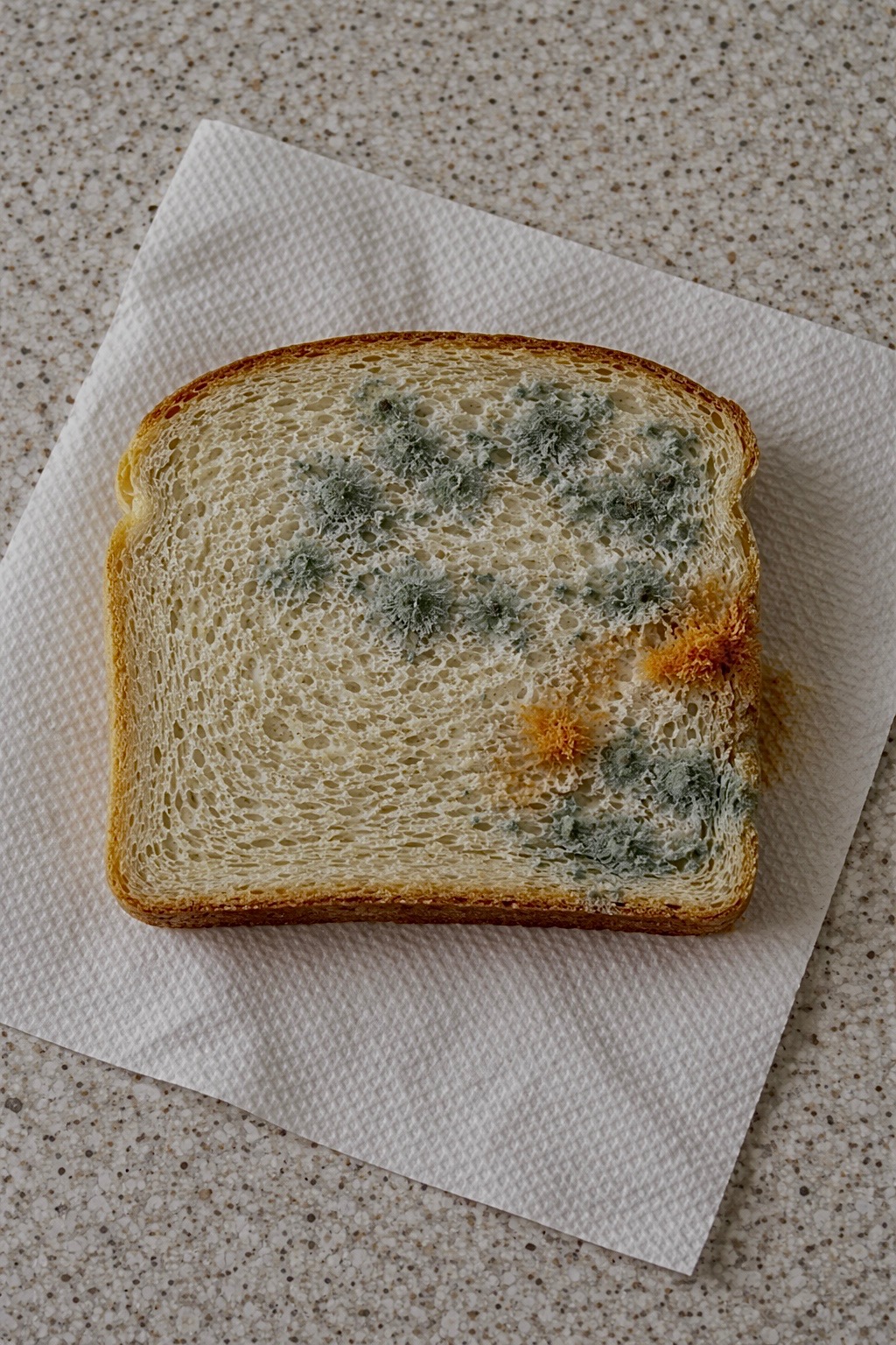
    I even did a test like this myself, leaving bread out for several days just to observe what happened to the mold (seen below).

    Somehow, this small organism allowed scientists to understand how living things, including humans, keep track of time using a biological clock.

    It did not seem too important at first. But as soon as they took it into their laboratory, it did something rather unusual. They placed the fungus in complete darkness, with no sunlight, not even a single outside signal. The fungus could not tell what time it was. Even then, it still grew in certain patterns (Castro-Longoria et al., 2010). It repeated in a consistent cycle, as though the organism already knew what a day looked like.

    This insight changed the perspective on time.

    Scientists such as Jay C. Dunlap and Jennifer J. Loros concluded that organisms do not just respond to time but keep an internal clock that runs in cycles (Castro-Longoria et al., 2010). Such a system came to be called a circadian rhythm, meaning “biological rhythm occurring every 24 hours.”

    Scientists later discovered that this clock runs through interactions among genes and proteins in a cycle. A vital part of this cycle includes a gene whose job is to create a certain type of protein named FRQ (frequency) (Baker et al., 2011).

    When the FRQ protein accumulates in the body, it stops being produced. When this protein degrades, production resumes. Through this process, a continuous cycle is formed, allowing time to be regulated. This is known as a negative feedback loop, where a system turns itself on and off (Baker et al., 2011).

    This system is not simple. However, there are other genes, which help the organism to sense light. These are collectively referred to as the “white collar” genes (WC-1 and WC-2). They work together in a cycle and are almost always around 24 hours in duration despite any change in the environment.

    Humans function with a similar system.

    The human brain has an intricate structure called the Suprachiasmatic Nucleus (SCN), which serves as the timekeeper for the organism, regulating physiological processes like sleep, hormone secretion, and body temperature. Just like in fungi, the circadian rhythm is regulated via protein-gene feedback loop mechanism.

    Circadian rhythms have been observed across different organisms, suggesting that the biological mechanism for keeping time is ubiquitous and therefore fundamental for any living thing (Aramayo & Selker, 2013).

    This was actually quite simple. Our internal clock had always been around.It was only when Neurospora crassa revealed itself that we realized its existence.

    References:

    Baker, C.L., Loros, J.J., & Dunlap, J.C. (2011). The circadian clock of Neurospora crassa.
    https://pmc.ncbi.nlm.nih.gov/articles/PMC3203324/

    Aramayo, R. & Selker, E.U. (2013). Neurospora crassa, a Model System for Epigenetics Research.
    https://pmc.ncbi.nlm.nih.gov/articles/PMC3783048/

    Case, M.E., Griffith, J., Dong, W., Tigner, I.L., Gaines, K., Jiang, J.C., Jazwinski, S.M., Arnold, J.,  (2005). The aging biological clock in Neurospora crassa.
    https://pmc.ncbi.nlm.nih.gov/articles/PMC4228622/

    Castro-Longoria, E., Ferry, M., Bartnicki-Garcia, S., Hasty, J., Brody, S. (2010). Circadian rhythms in Neurospora crassa: Dynamics of the clock component frequency visualized using a fluorescent reporter
    https://www.sciencedirect.com/science/article/pii/S1087184509002242

  • Circus Sea Slugs: How clown nudibranchs are like real-life Pokémon

    The Pokémon “Shellos” and its evolution “Gastrodon” (whose pink West Sea variant is one of my favorite Pokémon) resemble a sea slug. Don’t just take my word for it, though—in their Pokédex entries, Shellos and Gastrodon are referred to as the “Sea Slug Pokémon”. The Pokémon Goomy is referred to as a Mollusk Pokémon in many languages, and although sea slugs are a type of mollusk, the resemblance is less apparent. The team behind the games really didn’t have to change much from their real-life inspiration, though, as sea slugs are some of the most unreal-looking creatures in our oceans. As Helen Sullivan writes about sea slugs, which are also known as nudibranchs, in the Smithsonian Magazine, “It is really, really difficult not to compare them to Pokémon characters” (2024).

    Nudibranchs belong to the class Gastropoda, which falls under the phylum Mollusca (mollusks). The term sea slug is used to broadly refer to 11 different subgroups under Gastropoda that do not have hard outer shells. True nudibranchs account for only one of these subgroups, but the others are sometimes also referred to as nudibranchs for convenience (Davey & Willan, 2020). 

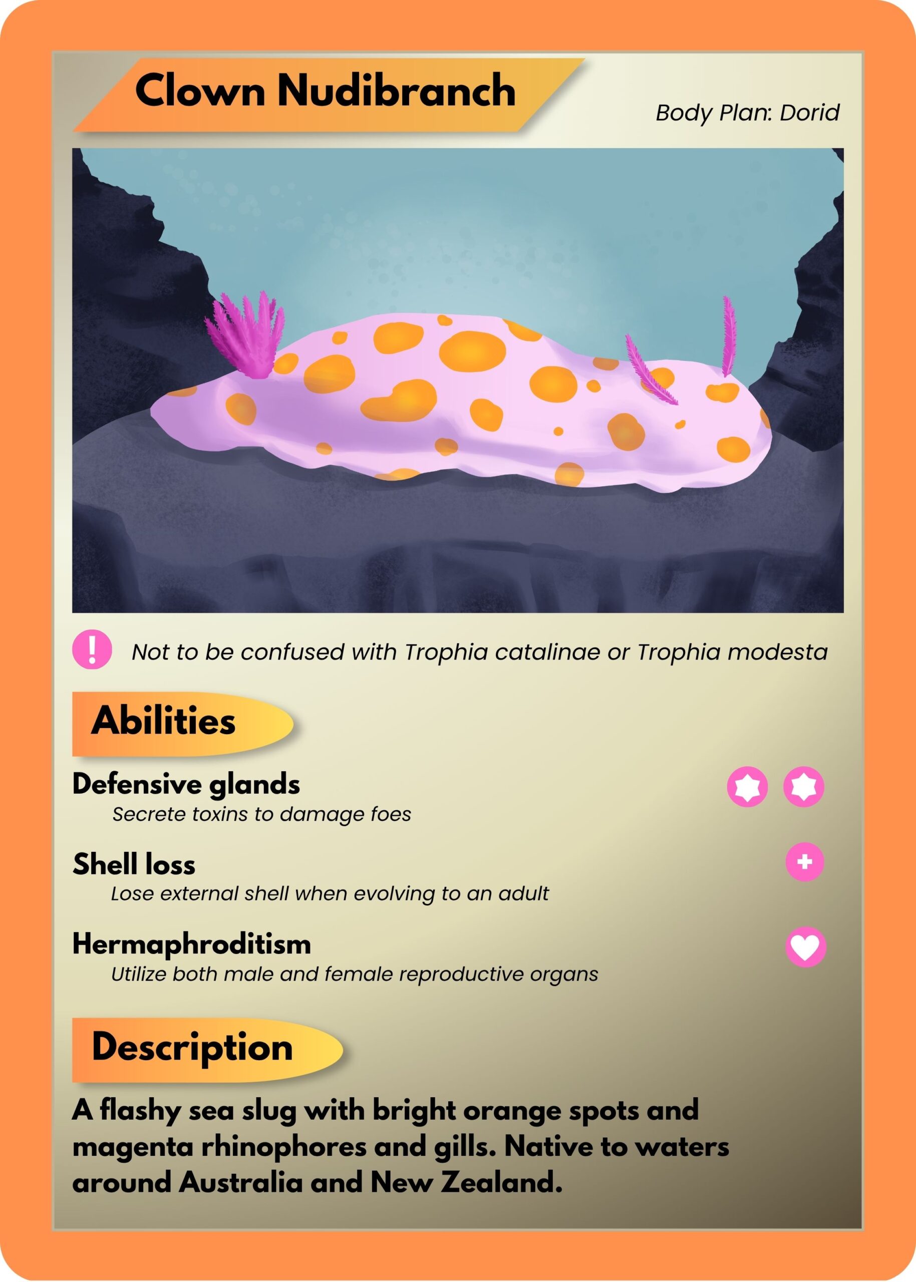
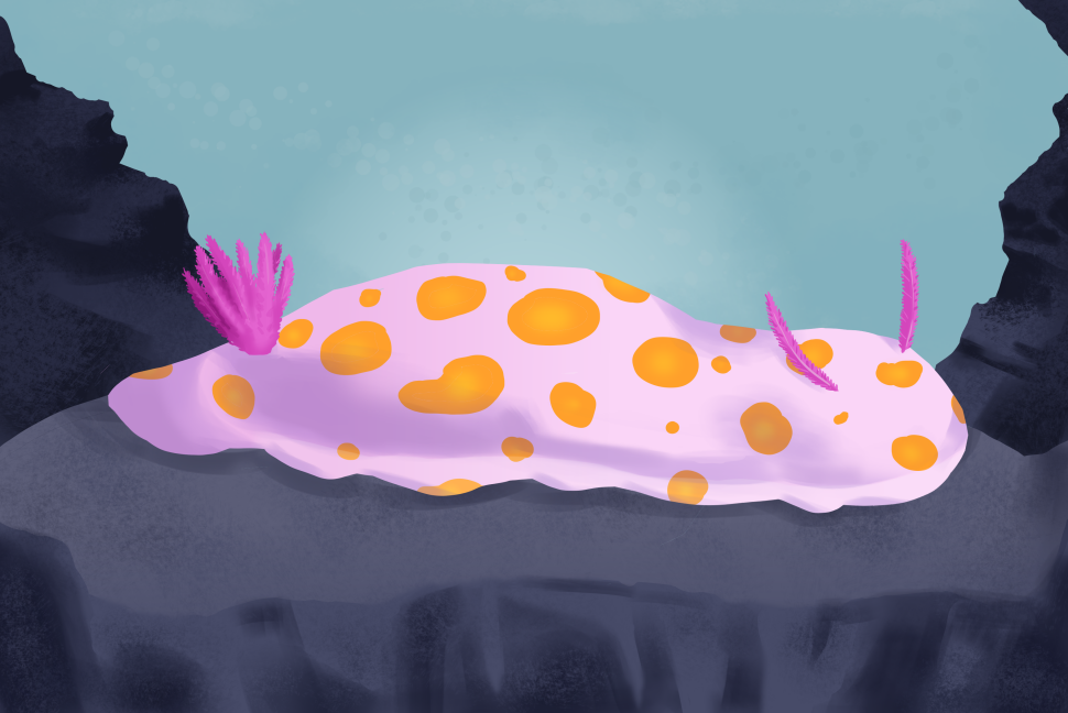
    When reading about species of nudibranch, one caught my eye for its name and bright orange coloration: the clown nudibranch. Even the name makes them sound like some sort of Pokémon! They are quite small—around 3 cm on average for adults—but their orange spots would probably make them hard to miss. The name, appearance, and distinctive characteristics of this creature gave me the idea to make a faux Pokémon card for the species, whose scientific name is Ceratosoma amoenum.

    Illustration/Graphic Design by Priyanka Singh

    Dorid

    Nudibranchs can have a few different body plans that differ in shape, gills, and more. C. amoenum has a dorid nudibranch body plan, which means it has two protrusions on its front end and a bundle of protrusions on its back end. The two protrusions that resemble antennae are called rhinophores while the bunch of protrusions on its backside are actually its gills! This distinctive characteristic is where nudibranchs get their name from. Nudibranch means “naked gills” (Sullivan, 2024).

    Not to be confused with…

    When researching for this article, I found that there are other nudibranch species with a similar appearance and common name as C. amoenum. Trophia catalinae also has a white body with orange spots, although its spots are raised (known as “tubercles”), and its rhinophores and gills are orange rather than magenta. It’s referred to by a similar name: “sea clown trophia” or simply “sea clown”, so you can see where the confusion might arise. Trophia modesta is a species that was recently distinguished from Trophia catalinae in a 2020 study, and it is commonly known as the “modest clown dorid” (Jung et al.). Unsurprisingly, it looks more similar to T. catalinae than it does to C. amoenum, although its body is more translucent than T. catalinae’s. Still, an amateur nudibranch enjoyer may get these species confused at first glance.

    Defensive glands

    Nudibranchs often contain chemical defenses within their bodies. A protective tissue called the mantle contains glands that secrete the toxins (Davey & Willan, 2020). Like other nudibranchs, our clown friend doesn’t produce these toxins on their own—they obtain them from their diet, which consists primarily of sponges (Winters et al., 2018). This chemical defense is critical for the soft, shell-less nudibranchs.

    Shell loss

    Speaking of not having a shell, both the English and Japanese name for Shellos mean “lost shell”: Karanakushi in Japanese, with kara meaning “shell” and nakushi being derived from nakushite (without) or nakusu (to lose); and “shell loss” (Shellos) in English (“Shellos”, 2026). The name origin references one of the nudibranch’s most defining characteristics. Gastropods have an external shell in their larval stage, although members of order Nudibranchia lose it before they reach adulthood, with C. amoenum being no exception (Davey & Willan, 2020). 

    Hermaphroditism

    As members of the Gastropoda class, sea slugs are hermaphrodites. Each individual has both male and female reproductive organs. I know what you may be wondering, but no, they can’t impregnate themselves, although they often donate and receive sperm simultaneously with another individual. Some nudibranchs even shed their penises after intercourse and gain a new one soon after (Sekizama et al., 2013). Talk about freaks of nature….

    I considered designing a creature based on the clown nudibranch for my fake Pokémon card, but why would I when the real thing looks like that? I’ll leave the creature designs to the Pokémon team for now. I hope you’ve come to see that sometimes reality can be stranger than the fiction it inspires, and that our world is full of amazing yet often overlooked creatures like the nudibranch.

    Illustration by Priyanka Singh

    References

    Davey, N., Willan, R.C. (2020). Super sea slugs: a guide to the sea slugs of New Zealand. (Version 1). National Institute of Water and Atmospheric Research (NIWA). 

    Gastrodon. (2026). Bulbapedia. Retrieved April 17, 2026, from https://pokemondb.net/pokedex/gastrodon 

    Jung, D., Gosliner, T.M., Choi, T., Kil, H., Chichvarkhin, A., Goddard, J.H.R., Valdés, A. (2020). The return of the clown: pseudocryptic speciation in the North Pacific clown nudibranch, Triopha catalinae (Cooper, 1863) sensu lato identified by integrative taxonomic approaches. Marine Biodiversity, 50, 84. doi.org/10.1007/s12526-020-01107-2 

    Sekizama, A., Seki, S., Tokuzato, M., Shiga, S., Nakashima, Y. (2013). Disposable penis and its replenishment in a simultaneous hermaphrodite. Biology Letters, 9(2). doi.org/10.1098/rsbl.2012. 1150 

    Shellos. (2026). Bulbapedia. Retrieved April 17, 2026, from https://pokemondb.net/pokedex/shellos 

    Sullivan, H. (2024, May). Dive Into the Exotic World of Nudibranchs, the Spectacular Slugs of the Sea. Smithsonian Magazine. www.smithsonianmag.com/science-nature/dive-exotic- world-nudibranchs-spectacular-slugs-sea-180984013/

    Winters, A.E., White, A.M., Dewi, A.S., Mudianta, I.W., Wilson, N.G., Forster, L.C., Garson, M.J., Cheney, K.L. (2018). Distribution of Defensive Metabolites in Nudibranch Molluscs. Journal of Chemical Ecology, 44, 384-396. doi.org/10.1007/s10886-018-0941-5 

  • Mandai Bird Paradise: Asia’s Biggest Aviary Sanctuary

    The Mandai Bird Paradise is the biggest bird conservatory in Asia, inaugurated back in May 2023. As one of the six pillars of the Mandai Wildlife Reserve, the incredible sanctuary covers over 42 acres of land on Mandai Island in Singapore. The reserve houses around 3,500 birds encompassing over 400 different species. Mandai Nature is the conservation arm of the Mandai Wildlife Group, which is dedicated to providing a haven for all these species through the Mandai Bird Paradise and to preserving Southeast Asia’s incredible biodiversity through conservation efforts, with a focus on averting species extinction (Temasek Trust, 2024). 

    Such a variety poses a rather obvious question: How? In order to answer this question, we must chart our way through the science behind conservation, biodiversity and rehabilitation!

    Conservation

    Conservation, as defined by the Merriam-Webster dictionary, is to carefully preserve and protect something. In a wildlife context, it is orchestrated management of natural resources to prevent exploitation, destruction, or neglect. Conservation efforts to preserve biodiversity are not just about protecting animals in isolation, but in our context, it is about the creation of an entire ecosystem where species can thrive naturally.

    At Mandai Bird Paradise, conservation takes an approach similar to this article on biodiversity conservation by diversity (Ramón et al., 2020). In-situ conservation focuses on protecting species in their natural habitats, whereas, ex-situ conservation, which is practiced at Mandai, provides controlled environments where vulnerable or endangered species can be bred, monitored, and protected from immediate threats such as habitat loss or poaching.

    The sanctuary also participates in international breeding programs and collaborates with global conservation organizations to ensure genetic diversity is maintained among species. This is critical because reduced genetic diversity due to issues such as environmental changes from global warming can make populations more susceptible to disease. Through these coordinated efforts, Mandai Bird Paradise becomes more than just a sanctuary, but a cornerstone in the Mandai Wildlife Group’s stand as an active contributor to global conservation science.


    Biodiversity

    Biodiversity refers to the variety of life in a particular habitat or ecosystem, encompassing differences in species, genetics, and ecological roles. The presence of over 400 bird species at Mandai Bird Paradise highlights not only the richness of different avian life present but also the complexity of maintaining such diversity within a single environment. This maintenance is done by separating the 42-acre land into around 10 unique habitats that each suit a certain group.

    Each species then gets to play its unique role in its ecosystem, from seed dispersal to pollination and pest control. By housing a wide array of species, the sanctuary effectively simulates interconnected ecological networks, allowing researchers and conservationists to better understand how these systems function.

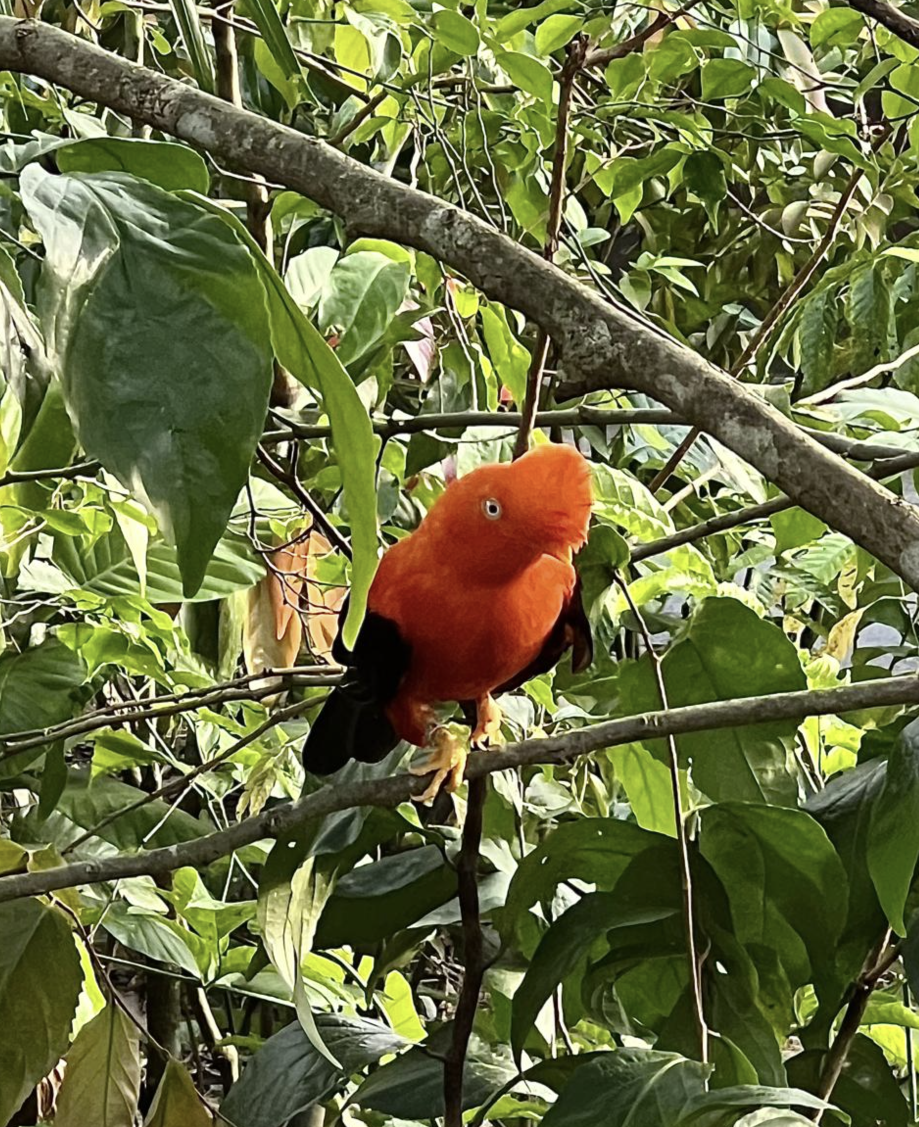
    Maintaining biodiversity in a controlled setting requires careful planning. Factors such as diet, climate conditions, nesting environments, and social groupings must all be tailored to meet the needs of different species. For instance, tropical birds such as the Andean cock-of-the-rock (yes, you read that right) require high humidity and dense vegetation, while others may need open spaces or specific nesting structures. This level of detail ensures that the birds are not only surviving but thriving in conditions that closely resemble their natural habitats.

    Andean cock-of-the-rock


    Rehabilitation

    Rehabilitation is another crucial component that involves the care and recovery of injured, displaced, or trafficked birds with the goal of reintroducing them into the wild whenever possible.

    Birds that arrive at rehabilitation centers often suffer from injuries caused by habitat destruction, illegal wildlife trade, or human interference. At Mandai, veterinary teams and specialists provide medical treatment, nutritional support, and behavioral conditioning to help these birds regain their strength and natural instincts.

    One of the key challenges in rehabilitation is ensuring that birds do not become overly dependent on human care. To address this, caretakers minimize human interaction and design enclosures that encourage natural behaviors such as foraging and flight. Only when a bird demonstrates sufficient recovery and independence is it considered for release. Even when release is not possible, rehabilitation still plays an important role. These birds may become part of educational programs, helping to raise awareness about conservation issues and inspiring visitors to take action in protecting wildlife.


    Mandai Bird Paradise – Sanctuary & Conservation Magic

    Mandai Bird Paradise is more than the visually stunning attraction you see through my lens, it is a living example of how science, conservation, and education can come together to protect our planet’s biodiversity. By integrating conservation strategies, maintaining diverse ecosystems, and supporting rehabilitation efforts, this sanctuary embodies what modern wildlife preservation should look like. In doing so, Mandai Bird Paradise not only safeguards thousands of birds but also inspires future generations like yours and mine to value and preserve the delicate balance of life on Earth.

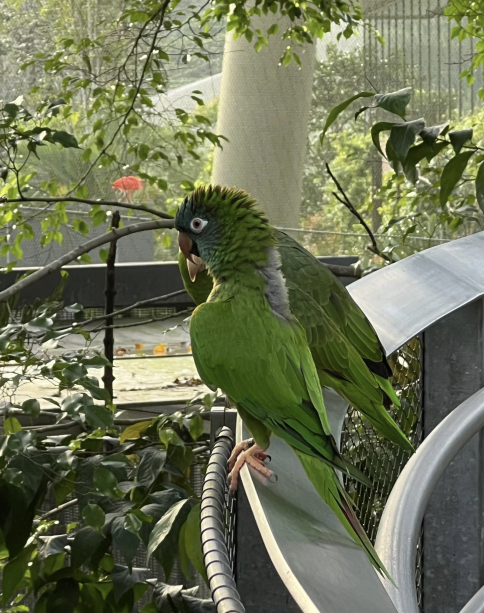
    Blue-crowned Parakeet

    Bits of My Time

    This reserve is one of the most beautiful places on Earth! Walking out into the Crimson Wetlands zone of the park felt like a scene straight out of a movie. There were colorful macaws soaring overhead, bright American flamingos dancing by the pond, Scarlet ibis carefully balancing on the hand rails and so much more. My exploration of the Mandai Reserve taught me so many fun facts about birds and this experience rekindled my love for all things nature. It truly gave me a fond memory to recall for the rest of my lifetime.

    References

    All Photography/Illustrations by Kristina Sarah Siju

    Mestanza-Ramón, C., Henkanaththegedara, S. M., Vásconez Duchicela, P., Vargas Tierras, Y., Sánchez Capa, M., Constante Mejía, D., Jimenez Gutierrez, M., Charco Guamán, M., & Mestanza Ramón, P. (2020). In-Situ and Ex-Situ Biodiversity Conservation in Ecuador: A Review of Policies, Actions and Challenges. Diversity, 12(8), 315. https://doi.org/10.3390/d12080315 

    Protecting Wildlife, Preserving The Future. (2023). Temasek Trust Revamp. https://www.temasektrust.org.sg/what-we-do/impact-hub/article-details/mandai-nature-impact-report-2023-2024

    Speiran S. I. M. (2025). The ‘Sanctuary Gap’: Reviewing the Research on Captive Wildlife Sanctuary Tourism. Animals : an open access journal from MDPI, 15(4), 496. https://doi.org/10.3390/ani15040496

  • Bouba? Kiki? Baby Chicks Associate Sounds With Shapes

    One of the chicks above is named “Bouba.” The other is named “Kiki.” Going just off of your gut intuition, which one is which?

    If you answered that the left chick is “Bouba” and the right chick is “Kiki”, many people agree with you, including speakers of over 25 languages, belonging to 9 language families and 11 writings systems—a surprisingly high number, given that these are purely nonsense words (Ćwiek et al., 2021). For some reason, “bouba” is obviously round, while “kiki” is obviously spiky.

    Perhaps even more astonishing is that, as discovered in a recent study by Maria Loconsole and colleagues at the University of Padova, baby chickens are among the population that agrees with you.

    What is the bouba-kiki effect?

    Most words have an arbitrary relationship to their meaning. Nothing about “cat” inherently refers to the feline domestic animal, and neither does 猫 (māo), बिल्ली (billi), gato, or قطة (qiṭṭa). But certain sounds seem to inherently have meaning.

    First described in 1947 by Wolfgang Kölher through the nonsense words “Maluma” and “Takete”, the “bouba-kiki” phenomenon in linguistics refers to this effect (Loconsole et al., 2026). “Maluma” and “bouba” feel inherently round, while “takete” and “kiki” feel inherently sharp and spiky. This effect is common across cultures, languages, and age: even infants as young as 4 months possess this sound mapping, where certain sounds seem to inherently correspond to certain shapes (Ozturk et al., 2013). This effect is even found (to a lesser extent) in blind individuals, though a period of sight seems necessary for the effect to occur (Piller et al., 2023).

    The most common explanation for the bouba-kiki effect is the way we say those syllables. When we say “bouba” and similar words, our mouths make a round shape; when we say “kiki” and similar words, our mouths make an angular shape. Our brains thus associate words that are “round” to pronounce with round shapes, and “angular” with angular shapes. Alternatively, the shape of the letters “b” and “o” are round, while the shape of the letters “k” and “i” are angular (Lockwood & Dingemanse, 2015).

    However, this hypothesis is challenged by the generic shape hypothesis, which states that humans form generic associations between certain sounds and meanings. This hypothesis was proposed in a study conducted by Ananya Passi and S. P. Arun in 2019, where the bouba-kiki effect was found with unpronounceable words, and an association was even found between clear tones and shape, where higher tones were associated with angular shapes. This indicates that humans potentially form general associations between sounds and shapes, regardless of the way those sounds can be pronounced. However, the reason why those associations might be formed remains unanswered, and these studies have been restricted to humans.

    Other animals have been shown to also experience some form of association between sound and another sense: for example, chimpanzees, tortoises, and dogs associate lower pitches with larger objects and higher pitches with smaller objects (Kalan et al., 2014; Korzeniowska et al., 2022; Loconsole et al., 2023). However, the bouba-kiki effect specifically has not been found to exist in other animals, even great apes, such as chimpanzees and a bonobo (Margiotoudi et al., 2019; Margiotoudi et al., 2022).

    Baby chicks?

    If the effect isn’t even found in our closest animal relatives, then where else can researchers look? In 2026, Loconsole and her colleagues decided to change tracks and look elsewhere. As surprising as their choice may seem, their pick of animals wasn’t arbitrary: chickens are precocial creatures, which means that they can be tested shortly after hatching, and chicks also share many cognitive mechanisms with human infants (Wood & Wood, 2021).

    In the first part of the study conducted by Loconsole and her colleagues, 3-day-old chicks were trained to go around a panel depicting a shape with both spiky and round edges, with food behind the panel acting as an incentive. During the experiment, the chicks were placed in front of two panels. One of the panels displayed a round shape, and the other one displayed a spiky shape, and neither of the panels concealed food. The researchers played a recording of a human voice saying either “bouba” or “kiki” to the enclosure, and they watched to see which panel the chicks would choose.

    In the second part of the experiment, 1-day-old chicks were introduced to an enclosure with a screen that once again showed a shape with both spiky and round edges, though there was no incentive. After the chicks grew accustomed to the screen, the researchers changed it up: like the first part of the experiment, the screen now showed two shapes side by side, with one jagged shape and one blobby shape. The researchers once again played either “bouba” or “kiki”, and they recorded which shape the chicks approached first, as well as which shape they spent more time exploring.

    The researchers found that the 3-day-old chicks significantly preferred the panel with the round shape when hearing “bouba”, and the panel with the angular shape when hearing “kiki”. They also found the same results with the 1-day-old chicks: when “bouba” was played, the chicks first approached the round shape significantly more and spent more time exploring the round shape. The reverse was true when “kiki” was played.

    What does this mean?

    The study shows that this bouba-kiki effect is not learned, but rather something inherent that’s present even in the earliest stages of life. One implication of this study is that this crossmodal association, or association between different senses, plays a role in how communication develops between animals. Animals such as chickens do not have language, but they still communicate through various calls and vocalizations. Although these vocalizations aren’t necessarily directly symbolic, having these innate associations may help with future development of more complex communication.

    Additionally, the findings of the study indicate that the bouba-kiki effect isn’t specific to humans or even mammals, but rather throughout all vertebrates. But how does this reconcile with past findings wherein the effect was not found in other great apes? After all, it’s rather unlikely that this association evolved separately in birds and in humans, but skipped our closest great ape relatives.

    According to Loconsole and colleagues, this difference may be explained by the difference in methodology between experiments, as well as the past experience and training of the research subjects. Kanzi, the bonobo who was tested for the bouba-kiki effect, was already trained in associating sounds and shapes and was capable of correctly matching English words to pictures, which may have played a role in his choices (Loconsole et al., 2026; Margiotoudi et al., 2022).

    So what do the findings of the study mean for the bouba-kiki effect and the development of human language? Does this mean that chickens are also capable of speech?

    No, not really. The bouba-kiki effect is only one example of the human ability of iconicity, or the capacity to form associations between form and meaning, argue Marcus Perlman and Bodo Winter in their commentary article in response to the 2026 study. In other words, the study reveals that the effect originates further back in the tree of life, but the origins of language lie elsewhere, in a broader framework. The bouba-kiki effect may reflect one way in which human language developed through associations between senses, but humans are able to form much more complex associations in language through a combination of speech, gestures, and visual signs. As such, researchers must look at other, broader areas for more clues in their hunt for the origins of language (Perlman & Winter, 2026).

    So overall, the chicks might agree with your naming of their illustrated compatriots, but unfortunately, they won’t be able to say as much in words.

    Want to learn more about the bouba-kiki effect? Check out our past article on the phenomenon!

    Writing and illustration by Joyce Ma


    References

    Ćwiek, A., Fuchs, S., Draxler, C., Asu, E. L., Dediu, D., Hiovain, K., Kawahara, S., Koutalidis, S., Krifka, M., Lippus, P., Lupyan, G., Oh, G. E., Paul, J., Petrone, C., Ridouane, R., Reiter, S., Schümchen, N., Szalontai, Á., Ünal-Logacev, Ö., Zeller, J., Perlman, M., Winter, B. (2021). The bouba/kiki effect is robust across cultures and writing systems. Philosophical Transactions of the Royal Society B Biological Sciences, 377(1841), 20200390. https://doi.org/10.1098/rstb.2020.0390

    Kalan, A. K., Mundry, R., & Boesch, C. (2015). Wild chimpanzees modify food call structure with respect to tree size for a particular fruit species. Animal Behaviour, 101, 1–9. https://doi.org/10.1016/j.anbehav.2014.12.011

    Korzeniowska, A. T., Simner, J., Root-Gutteridge, H., & Reby, D. (2022). High-pitch sounds small for domestic dogs: Abstract crossmodal correspondences between auditory pitch and visual size. Royal Society Open Science, 9(2), 211647. https://doi.org/10.1098/rsos.211647

    Lockwood, G., & Dingemanse, M. (2015). Iconicity in the lab: A review of behavioral, developmental, and neuroimaging research into sound-symbolism. Frontiers in Psychology, 6, 1246. https://doi.org/10.3389/fpsyg.2015.01246

    Loconsole, M., Benavides-Varela, S., & Regolin, L. (2026). Matching sounds to shapes: Evidence of the bouba-kiki effect in naïve baby chicks. Science, 391(6787), 836–839. https://doi.org/10.1126/science.adq7188

    Loconsole, M., Stancher, G., & Versace, E. (2023). Crossmodal association between visual and acoustic cues in a tortoise (Testudo hermanni). Biology Letters, 19(7), 20230265. https://doi.org/10.1098/rsbl.2023.0265

    Margiotoudi, K., Allritz, M., Bohn, M., & Pulvermüller, F. (2019). Sound symbolic congruency detection in humans but not in great apes. Scientific Reports, 9(1), 12705. https://doi.org/10.1038/s41598-019-49101-4

    Margiotoudi, K., Bohn, M., Schwob, N., Taglialatela, J., Pulvermüller, F., Epping, A., Schweller, K., & Allritz, M. (2022). Bo-NO-bouba-kiki: Picture-word mapping but no spontaneous sound symbolic speech-shape mapping in a language trained bonobo. Proceedings of the Royal Society B Biological Sciences, 289(1968), 20211717. https://doi.org/10.1098/rspb.2021.1717

    Ozturk, O., Krehm, M., & Vouloumanos, A. (2012). Sound symbolism in infancy: Evidence for sound–shape cross-modal correspondences in 4-month-olds. Journal of Experimental Child Psychology, 114(2), 173–186. https://doi.org/10.1016/j.jecp.2012.05.004

    Passi, A., & Arun, S. P. (2022). The Bouba–Kiki effect is predicted by sound properties but not speech properties. Attention Perception & Psychophysics, 86(3), 976–990. https://doi.org/10.3758/s13414-022-02619-8

    Perlman, M., & Winter, B. (2026). In search of meaning. Science, 391(6787), 762–763. https://doi.org/10.1126/science.aee8641

    Piller, S., Senna, I., & Ernst, M. O. (2023). Visual experience shapes the Bouba-Kiki effect and the size-weight illusion upon sight restoration from congenital blindness. Scientific Reports, 13(1), 11435. https://doi.org/10.1038/s41598-023-38486-y

    Wood, S. M. W., & Wood, J. N. (2021). One-shot object parsing in newborn chicks. Journal of Experimental Psychology General, 150(11), 2408–2420. https://doi.org/10.1037/xge0001043

  • Why Is Math So Hated?

    “What’s your favorite subject?”

    “I’m not sure… just definitely not math. What’s yours?”

    “Oh, funny story…”

    Ah yes, math, the subject notorious for being one of the hardest subjects in school, with “math anxiety” being the term coined for the fear of math. If you’re reading this, you’ve most likely been through the agony of a difficult math class or teacher that just didn’t understand you. As a math major and someone who loved math as a child (but also had their share of math difficulties), I sought out to understand why math is so hated, and if there is anything we can do to change that. After all, I was able to see firsthand how cool math is and my hope is that more and more people will be able to as well.

    How Math is Taught in School

    Many students point to the difficulty of math when asked why they dislike it, as shown from this table, where math being hard was listed as a reason almost twice as much as it being boring (Stodolsky et al., 1991). Therefore, we should look to how math is taught in school to see why students are struggling so much. In general, many math educators teach in a more computationally-focused way, prioritizing students’ abilities to memorize concepts and algorithms to be able to complete a problem set, rather than letting them discover the math on their own or letting the students know the “why” behind what they are doing (Stodolsky et al., 1991). This was certainly my experience in some of my classes, where when asked why something works the teacher would just say “don’t worry about it.” This can cause students to be confused on how to follow the steps given to them to solve a problem or give them difficulty when trying to memorize them for a test. If students knew all of the derivations to the formulas they were using, I think they would be more likely to remember it and boost their problem-solving skills than if they regurgitate a step-by-step process given to them.

    Gender Stereotypes Regarding Math Ability

    If you’re a girl, you may have heard it at least once in your life: “You’re a girl? You can’t possibly be good at math or science, you belong in humanities (or the kitchen).” Or perhaps, you’ll be the only girl, or one of a few, in some advanced STEM class. One study was held testing children in a few different categories: math performance, math anxiety, and test anxiety, collecting data over different grades for boys versus girls. The study found that higher math anxiety had more of a negative effect on girls than for boys (Erturan & Jansen, 2015). This is likely due to the aforementioned  stereotypes that boys are better than girls at STEM related subjects such as math, putting more pressure on girls when testing. Even if they might know the content, these stereotypes can put doubt into girls’ minds about whether or not they are capable of doing well in math.

    Intergenerational Effects on Math Anxiety

    Math anxiety has existed for a long time, not just in this generation, but in those of our parents and grandparents as well. As a result, it makes sense to consider the possibility that it gets passed down from generation to generation, not necessarily through genetics, but through parents’ attitudes towards math rubbing off on their children. Another study on math anxiety investigated the role that parents played pertaining to their children’s levels of math anxiety, by testing students’ reading and math performance and giving parents questionnaires to determine their levels of math anxiety. The study revealed that there was a negative correlation between a parent’s math anxiety and their child’s performance, but only when the parent was significantly involved with helping the child with their homework (Maloney et al., 2015). It is speculated that the cause of this is the parents letting out their own negativity towards the subject to the child, expressing their frustration which caused their children to also have higher levels of math anxiety and do poorer on their tests.

    What To Do?

    It is clear there are many outside factors that can cause a child to dislike math, so  how do we combat this? Even if we don’t like math that much ourselves, we can try our best not to project that mindset onto young minds and instead, encourage students to develop a growth mindset (Mendoza, 2025). With some work, it would also be beneficial to make a few changes to the way math is taught in school by making learning more interactive and prioritizing understanding over memorization. And hopefully, maybe one day, math will not be considered the “hated” subject but simply just a subject that more people enjoy.


    References

    Erturan, S., & Jansen, B. (2015). An investigation of boys’ and girls’ emotional experience of math, their math performance, and the relation between these variables. European Journal of Psychology of Education, 30(4), 421–435. https://www.jstor.org/stable/24763249

    Maloney, E. A., Ramirez, G., Gunderson, E. A., Levine, S. C., & Beilock, S. L. (2015). Intergenerational Effects of Parents’ Math Anxiety on Children’s Math Achievement and Anxiety. Psychological Science, 26(9), 1480–1488. https://www.jstor.org/stable/24544088

    Mendoza, K. (2025, February 28). 9 Strategies to Help Students Love Math – Oxford Learning. Oxford Learning. https://oxfordlearning.com/9-strategies-to-help-students-love-math/

    Stodolsky, S. S., Salk, S., & Glaessner, B. (1991). Student Views about Learning Math and Social Studies. American Educational Research Journal, 28(1), 89. https://doi.org/10.2307/1162880

  • The Hidden World of the Piano Action

    The cover image was taken by the author.

    In my humble opinion, the best way to unwind after a long, arduous day is to pour out all your stresses onto the beautiful white and black keys of a grand piano.

    (Of course, that’s a little hard to do if you’ve never learned to play… I digress, though.)

    During my time at Georgia Tech, I’d find myself sitting at the piano located in the West Village Dining Hall almost every night – and while I wouldn’t say the piano there is especially well maintained, it still produces a sound that’s pleasant to my ears. In the end, isn’t that all that matters?

    “Enough with the preamble! What’s the point of all these ramblings?” You might be asking. Well, to answer that hypothetical question – I often think about the acoustics of a piano. That, of course, naturally leads to how it works… and believe it or not, there happens to be a surprising amount of physics under the (metaphorical and literal) hood of the instrument!

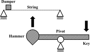
    The story of the piano begins with the clavichord, developed in the 14th century. The clavichord action was as simple as could be – a key lever with a hammer on one end. After the key is depressed, the hammer is immediately lifted until it hits the string, remaining in contact until the key is released (Russo & Robles-Linares, 2020).

    Figure 2 Source: (Russo & Robles-Linares, 2020)

    While this action enables volume dynamics, vibrato, and control over the attack and duration of one’s sounds, there remained one large problem: the string would be damped for as long as the key was held down.

    The harpsichord attempted to address this issue, while sacrificing some of the finer benefits of the clavichord.

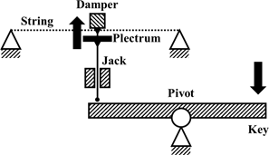
    For its action, when a key is pressed, the jack is lifted along with the plectrum and damper, the latter immediately releasing the string. Right after, the plectrum hits the string, generating sound. At some point, the jack meets a rail up above that stops its motion, allowing the string to resonate for as long as the key is held down. When the key is finally let go, the jack falls such that its plectrum does not hit the string a second time (Russo & Robles-Linares, 2020).

    Figure 3 Source: (Russo & Robles-Linares, 2020)

    Even though this solves the major issue with the clavichord, the harpsichord, in turn, has almost no volume dynamics, nor can vibrato be performed with it!

    That takes us to the mid-17th century, where Bartolomeo Cristofori would invent what was known as the fortepiano – named because of its ability to play forte (loud) and piano (soft).

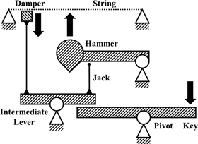
    The action of the fortepiano is far more complex than both the clavichord and the harpsichord. When the key is first pressed, it lifts an intermediate lever; a damper connected to that lever releases the string. After a small period of time, the jack on the intermediate lever pushes the hammer up. The hammer lifts off the jack halfway to the string and uses its existing momentum to hit the string.

    If the key was immediately released, the string is immediately damped, making a staccato (short) sound, as the mechanism returns to its resting position. If the key is held down, the string is undamped and free to resonate; the hammer falls back down onto the jack. After the key is released, the hammer returns to its resting position and the string is damped, muting the sound (Russo & Robles-Linares, 2020).

    Figure 4 Source: (Russo & Robles-Linares, 2020)

    In essence, the fortepiano was the combination of the good of both the clavichord and harpsichord – dynamics and vibrato, along with volume and an ability to sustain sound. In many ways, the fortepiano is similar to the grand pianos of today, but with a few key differences…

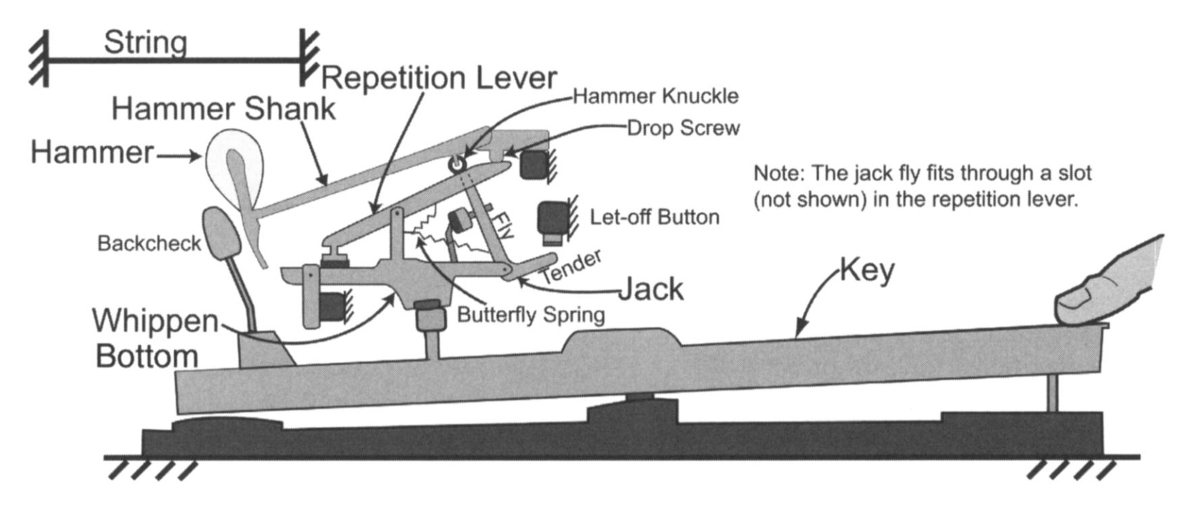
    Figure 5 Source: (Brent et al., 2011)

    …Quite a lot of differences, in fact. From a first glance, this diagram seems far removed from the likes of the fortepiano, but most of the underlying theory remains the same.

    To put it simply, the goal of the grand piano action is to let the hammer hit the string to create vibration – but, most importantly, it should be fast enough that it doesn’t damp the string. Efforts to reduce that time have resulted in the complex system seen in Figure 5… each part of that mechanism plays its own role in giving as much control to the pianist as possible (Brent et al., 2011).

    Control is an important part of playing the piano, in my own experience. The difference between a grand piano and an electronic MIDI keyboard is like night and day. Even the smallest, involuntary actions when playing a grand result in minor yet noticeable changes – and the best of the best (which, unfortunately, doesn’t include me) are able to manipulate each and every facet of control given to them.

    As we close the hood on the fascinating world of the piano action, I hope you’ve gained a proper appreciation for the instrument! The fact that we’ve been able to manufacture such a sleek design, fitting in all the complex mechanisms into such a small area, all while ensuring it works to perfection… it’s an engineering miracle, to be honest.

    When I inevitably return to that piano in West Village, I’ll surely be thinking about the action that I’m invoking with every press.

    Reference List

    Brent, G. R., Yu, B., Grijalva, R., & Awtar, S. (2011). Characterizing the Feel of the Piano Action. Computer Music Journal, 35(1), 43–57. JSTOR. https://www.jstor.org/stable/41241706

    Russo, M., & Robles-Linares, J. A.  (2020). A Brief History of Piano Action Mechanisms. Advances in Historical Studies, 09(05), 312–329. https://doi.org/10.4236/ahs.2020.95024

  • Organoids: What are They and Why They’re Amazing

    I’m willing to bet you’ve never heard of Organoids. No worries if so, they’re a pretty niche part of biomedical sciences. This is such a shame however as they’re having a great impact on research and our world. Organoids are often defined as tissue samples embedded into 3D extracellular matrix (ECM) gels or clonal derivatives of cells grown without mesenchyme and derived from stem cells. Don’t worry if your eyes have already glazed over! Mine did too the first time I read that.

    In simpler terms, they are the growing of cells, taken from a patient, in a nutrient rich gel. While the field might seem niche at first, it has a long history and the potential to impact millions.

    Before Organoids, most human physiological research relied on in vitro 2D cell culture and animal models (O’Connell & Winter, 2019). These often fail at clinical trials due to oversimplification of or incompatibility with human systems. At the time, cell cultures were limited to growing single layers of cells in a petri dish with none of the 3D complexities of real biological systems. This all changed with Ross Harrison’s “Hanging drop Cell Cultures” in 1906 (Simian and Bissell, 2016). By creating a 2D cell culture and turning the petri dish upside down, gravity would cause the cells to clump together and form a cohesive 3D spheroid of cells that could intermingle and interact like normal. Think about that! 3D structures could form by simply turning them upside down with no other interference. This was the start of research into organoids which offered a new way to look directly at human biology without risking patient or animal lives.

    One key breakthrough was the discovery of 3D extracellular matrices first used in 1963 (Simian and Bissell, 2016). The gel provided a medium for natural scaffolding of cells rather than relying on gravity. This allowed for more complex 3D shapes and allowed for more types of organoids that more closely resembled complex real life systems. The gel also aided in providing the cells with nutrients required to keep them alive. Hence the “nutrient rich gel” from the definition above.

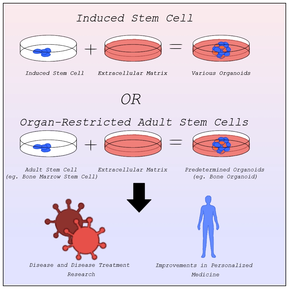
    Exploration into this new field was rather slow however. The human body is composed of roughly 200 distinct types of cells, each of which specialize in a different function such as blood or nerve cells. Despite this, organoids mainly rely on only two types of cells, pluripotent stem cells and organ-restricted adult stem cells (Bredenoord et al., 2017). Stem cells are unspecified types of cells, yet to differentiate, with the ability to become any type of specified cell. They often serve to repair the human body as they can replace any damaged cells no matter the type. This ability makes them highly valuable to organoids trying to model systems that rely on the cooperation of multiple cell types.

    Before the discovery of induced stem cells in 2006 by Dr. Shinya Yamanaka at Kyoto University, organoids were made from pluripotent stem cells as organ-restricted adult stem cells can be hard to access and are restricted to becoming cell types of their tissue of origin most commonly found in embryos (Agarwal et al., 2024). The line of when a zygote becomes a person, however, is blurry, raising many ethical concerns about the desecration of a person’s body. Induced stem cells allow for the change of specialized cells back into stem cells. Organoids can now be created from any tissue sample, entirely limiting the need for embryonic stem cells outside of studying early organ development (Frans Schutgens & Clevers, 2019).

    With better ways to develop organoids, scientists quickly realized their potential. Organoids offer unparalleled advancement in disease research and treatment, personalized medicine, and so much more that we simply don’t have time for here.

    Image by Alexandra Marshall

    Disease Research and Treatment
    One pivotal area of applications is understanding the development of diseases and disease treatment in humans. The lack of 3D structure in in vitro models lead to limited complexity and physiological relevance when studying diseases. Faulty predictions based on these studies can lead to treatments failing, with approximately 90% of clinical study drug candidates failing clinical trials and drug approval (Sun et al., 2022).

    Organoid models have none of these limitations. In 2013, the first human condition modeled using organoids, cystic fibrosis, was published. The model had “excellent predictive value” and became the first successful personalized treatment method for cystic fibrosis patients (Lancaster and Huch, 2019). The modeling of other diseases using organoids quickly followed from genetic diseases like Polycystic kidney disease and Alagille syndrome, to infectious diseases like Covid-19 and the Zika virus, to neurological diseases like Alzheimer’s and Parkinson’s, to multitudes of cancers, and more (Xu et al., 2018). The ability for organoids to be made of any human system means they can model any disease regardless of which systems they afflict.

    Organoids can be also grown co-culturally with bacteria allowing for bacterial infections and the diseases that arise from them to also be modeled and researched with organoids (Schutgens and Clevers, 2020). This has allowed for better understanding of bacteria like Helicobacter Pylori and Salmonella, aiding in detection and cures. As mentioned above, treatments can then be tested on these organoid systems without risking human lives or animal cruelty.

    Respiratory Syncytial Virus (RSV), a disease that is being modeled in airway organoids, causes around 66,000–199,000 deaths among children annually (Schutgens & Clevers, 2020). This is just one of the many diseases for which scientists are now better equipped to find viable treatments. Organoids have already saved millions of lives and their potential for future disease treatment has the potential to millions more.

    Personalized Medicine
    While these treatments work for many people, genetic and biological differences can impact treatment effectiveness and tolerance with some even leading to negative side effects (Breen, 2021). In such situations, personalized medicine that is specialized to fit these differences might be necessary. Personalized medicine, sometimes called precision medicine, challenges the idea that treatments that pass general trials will work for everyone, instead working to construct a treatment plan with a specific patient in mind best suited to their physiology.

    The idea of personalized medicine has been around for thousands of years, using one’s family’s history of diseases to diagnose the likelihood of that person having said disease. In the late 1900s the idea of evidence based medicine, the implementation of explicit reasoning based on a specific patient’s data, started being common practice when prescribing a patient (Visvikis-Siest et al., 2020). This opened the door for personalized medicine in modern science (Visvikis-Siest et al., 2020). This data was largely based on patients’ reactions to other treatments and family history however limited its effectiveness. The development of molecular biology and human genome mapping allows for a direct look at a patient’s physiology, aiding medical professionals in determining the biological reasons behind potential reactions to treatments before administration limiting adverse reactions.

    Organoids have greatly furthered this field. As mentioned above, organoids are made from a patient’s induced stem cells. This means that the organoid culture into a structure based on the patient’s DNA, containing all the same biomarkers and physiology that the patient would have at that level. Where other treatments could have varying effects from the tested population, organoids allow medical professionals to see the exact effects a treatment plan would have on the patient. The use of stem cells also allows organoids to grow much quicker than other models. This allows for quick and accurate testing of the effects of medication on a patient. One study published in Science involving gastrointestinal cancer therapies found that organoids would correctly detect when drugs would fail 100% of the time, and when the drug would succeed 88% percent of the time (Vlachogiannis et al., 2018). Once again, because of the diversity of systems that organoids can model, organoids can be used to model many different personalized medicine plans allowing for the variety of treatment testing needed to find the most optimal plan.

    Conclusion
    Organoids have had a monumental, yet widely unknown, impact on a wide variety of disease advancement and treatment developments and personalized medicine. These structures can then be used to model human tissues and organs with much higher accuracy and complexity than previously used non-human or in vitro models.

    Scientists use them to model disease development in humans and treatments designed for humans. This allows for a better understanding of the diseases and more effective treatments with fewer errors. They are also used to limit the risk of negative side effects from non-personalized medicine by furthering personalized medicine through the accuracy and speedy testing of many different treatment options.
    With continued and careful development of organoids, disease understanding and treatments, personalized medicine and so much more can continue to be improved, saving millions of lives.

    ————————————————————————————————-

    Work Cited:
    Agarwal, S., Saha, L., Sohani, A., Karri, U., Gor, R., & Ramalingam, S. (2024). The importance of niche in therapeutic strategies targeting normal and cancer stem cells. Cancer Stem Cells and Signaling Pathways, 15–33. https://doi.org/10.1016/b978-0-443-13212-4.00031-3

    Bredenoord, A. L., Clevers, H., & Knoblich, J. A. (2017). Human tissues in a dish: The research and ethical implications of organoid technology. Science, 355(6322). https://doi.org/10.1126/science.aaf9414

    Frans Schutgens, & Clevers, H. (2019). Human Organoids: Tools for Understanding Biology and Treating Diseases. Annual Review of Pathology Mechanisms of Disease, 15(1), 211–234. https://doi.org/10.1146/annurev-pathmechdis-012419-032611

    Generation and Cultivation of 3D Cell Models | ibidi. (2024). Ibidi. https://ibidi.com/content/1037-generation-and-cultivation-of-3d-cell-models

    Lancaster, M. A., & Huch, M. (2019). Disease modelling in human organoids. Disease Models & Mechanisms, 12(7). https://doi.org/10.1242/dmm.039347

    O’Connell, L., & Winter, D. C. (2019). Organoids: Past Learning and Future Directions. Stem Cells and Development, 29(5), 281–289.https://doi.org/10.1089/scd.2019.0227

    Schutgens, F., & Clevers, H. (2020). Human Organoids: Tools for Understanding Biology and Treating Diseases. Annual Review of Pathology: Mechanisms of Disease, 15(1), 211–234. https://doi.org/10.1146/annurev-pathmechdis-012419-032611

    ‌Simian, M., & Bissell, M. J. (2016). Organoids: A historical perspective of thinking in three dimensions. The Journal of Cell Biology, 216(1), 31–40. https://doi.org/10.1083/jcb.201610056

    Sun, D., Gao, W., Hu, H., & Zhou, S. (2022). Why 90% of clinical drug development fails and how to improve it? Acta Pharmaceutica Sinica B, 12(7), 3049–3062. https://doi.org/10.1016/j.apsb.2022.02.002

    Visvikis-Siest, S., Theodoridou, D., Kontoe, M.-S., Kumar, S., & Marschler, M. (2020). Milestones in Personalized Medicine: From the Ancient Time to Nowadays—the Provocation of COVID-19. Frontiers in Genetics, 11. https://doi.org/10.3389/fgene.2020.569175

    Vlachogiannis, G., Hedayat, S., Vatsiou, A., Jamin, Y., Fernández-Mateos, J., Khan, K., Lampis, A., Eason, K., Huntingford, I., Burke, R., Rata, M., Koh, D.-M., Tunariu, N., Collins, D., Hulkki-Wilson, S., Ragulan, C., Spiteri, I., Moorcraft, S. Y., Chau, I., & Rao, S. (2018). Patient-derived organoids model treatment response of metastatic gastrointestinal cancers. Science, 359(6378), 920–926. https://doi.org/10.1126/science.aao2774

    Xu, H., Jiao, Y., Qin, S., Zhao, W., Chu, Q., & Wu, K. (2018). Organoid technology in disease modelling, drug development, personalized treatment and regeneration medicine. Experimental Hematology and Oncology, 7(1). https://doi.org/10.1186/s40164-018-0122-9